
Related Manuals for GE VOLUSON 730Pro


Summary of Contents for GE VOLUSON 730Pro
- Page 1 ® ® GE Medical Systems Kretztechnik GmbH & Co OHG...
- Page 2 GE Medical Systems Kretz Ultrasound Basic User Manual 105831 Revision 0 ® VOLUSON 730Pro 0366 © Copyright 2002 by GE Medical Systems - Kretztechnik GmbH & Co OHG User Documentation...
- Page 3 Revision History Table i-1: Reason for Change DATE REASON FOR CHANGE Rev.0 July 31, 2002 Initial Release Table i-2: List of effective Pages CHAPTER- / PAGE NUMBER REV # CHAPTER- / PAGE NUMBER REV # Cover Chapter 11 - Volume Mode Revision History Chapter 12 - Utilities Contents...
- Page 4 This page intentionally left blank. ® Voluson 730Pro - Operation Manual 105831 Rev. 0...
-
Page 5: Table Of Contents
CONTENTS REVISION HISTORY………………………………………………………………………i-1 GENERAL...................... 1-2 SAFETY ......................2-2 Important Instructions for Safety ..................2-2 Electric installation......................2-3 Symbols Used ........................2-3 Remarks for Safe Use ......................2-5 Environmental Conditions for Operation ................. 2-6 Instruction for Use....................... 2-6 Biopsy Lines ......................... 2-7 ECG preamplifier (MAN) .................... - Page 6 CONTENTS System Assembly........................3-6 3.3.1 Basic System........................3-6 3.3.2 Optional Peripheral Devices ....................3-6 3.3.3 Optional Modules......................3-7 3.3.4 Position of Display Annotation..................3-7 Concept of Operation......................3-8 3.4.1 Changing of Menus......................3-9 3.4.2 Layout of the 2D-Mode Main Menu................3-9 3.4.3 Control Panel .........................3-11 3.4.4 Hard keys ........................3-12 3.4.4.1 Function of the Trackball at Diverse Dialog Pages ..........3-18 3.4.4.2...
- Page 7 CONTENTS 2D MODE ....................... 5-2 2D Main Menu ........................5-2 2D Mode Depth ........................5-4 2D Image Angle........................5-4 2D Gain..........................5-5 TGC Slider Controls......................5-5 2D Automatic Optimization....................5-6 Transmit Power ........................5-6 Transmitter Focus ....................... 5-7 Receiver Frequency Range ....................5-7 5.9.1 Harmonic Imaging ......................
- Page 8 CONTENTS M MODE ......................6-2 M Main Menu........................6-2 6.1.1 Principle ...........................6-3 M Operation .........................6-4 6.2.1 Cursor Position.........................6-4 6.2.2 Activation of M Mode......................6-4 6.2.3 Sweep Speed ........................6-5 6.2.4 Invert ..........................6-5 6.2.5 Frequency.........................6-5 6.2.6 TGC Slide Controls......................6-5 6.2.7 M Mode Depth.........................6-5 6.2.8 M Gain Control ........................6-6 6.2.9 M Cineloop ........................6-6...
- Page 9 CONTENTS SPECTRAL DOPPLER MODE ..............7-2 PW Mode (Pulsed Wave Doppler) ..................7-2 7.1.1 PW Main Menu ....................... 7-3 7.1.2 PW Operation ........................7-4 7.1.2.1 Gate Position and Gate Width ..................7-4 7.1.2.2 Activation of PW Mode....................7-5 7.1.2.3 PW Gain Control ......................7-5 7.1.2.4 PW Automatic Optimization ..................
- Page 10 CONTENTS CFM MODE (COLOR FLOW MODE)............8-2 CFM Main Menu .........................8-2 CFM Operation........................8-3 8.2.1 CFM Box Position and CFM Box Size................8-4 8.2.2 CFM Gain Control ......................8-4 8.2.3 Quality..........................8-5 8.2.4 WMF..........................8-5 8.2.5 Velocity Range (PRF)......................8-6 8.2.6 Frequency.........................8-6 8.2.7 Invert ..........................8-7 8.2.8 2D + 2D/C........................8-7 8.2.9 Threshold .........................8-7...
- Page 11 CONTENTS PD MODE (POWER-DOPPLER MODE)............9-2 PD Main Menu........................9-3 PD Operation ........................9-4 9.2.1 PD Box Position and PD Box Size .................. 9-4 9.2.2 PD Gain Control ......................9-5 9.2.3 Quality ..........................9-5 9.2.4 Frequency ........................9-6 9.2.5 WMF..........................9-6 9.2.6 Velocity Range (PRF)......................
- Page 12 CONTENTS VOLUME MODE................... 11-3 11.1 Volume Acquisition with Volume Probes ................11-4 11.1.1 Principle of Volume Acquisition ...................11-5 11.1.2 Principal Scanning Modes .....................11-5 11.1.3 What is Interactive 3D Image Rendering ? ..............11-6 11.1.3.1 What is Interactive ? ....................11-6 11.1.4 Image Orientation (All Acquisition Modes) ..............11-7 11.1.5 The Render Box ......................11-9 11.1.6...
- Page 13 CONTENTS 11.5 Real Time 4D Acquisition ....................11-52 11.5.1 Possible Display Adjustment Before Starting Acquisition.......... 11-54 11.5.2 Real Time 4D Acquisition During Active High Resolution Zoom ......11-55 11.5.2.1 Display of sectional Planes..................11-56 11.5.2.2 Display of REF-Image.................... 11-57 11.5.2.3 Display of ROI 4D....................
- Page 14 CONTENTS BASIC MEASUREMENTS ................13-2 13.1 Basic Operations ........................13-3 13.2 Distance Measurements.....................13-4 13.2.1 2D Distance Measurement .....................13-4 13.2.2 M Distance Measurement ....................13-5 13.3 Area Measurements ......................13-5 13.3.1 Measurement using Ellipse ....................13-6 13.3.2 Measurement using Trace ....................13-6 13.4 Volume Measurements ......................13-7 13.4.1 3 Distance Measurement....................13-7 13.4.2...
- Page 15 CONTENTS CALCULATIONS AND PATIENT REPORTS..........14-3 14.1 Basic Calculation Functionality..................14-3 14.2 Basic Patient Report Functions ..................14-5 14.2.1 To view a Patient Report ....................14-5 14.2.2 To save / send a Patient Report..................14-6 14.2.3 To edit a Patient Report ....................14-7 14.2.4 To view previous Reports....................
- Page 16 CONTENTS 14.7.4.7 Ao/LA ........................14-33 14.7.4.7.1 To measure all Items at a Time..............14-33 14.7.4.7.2 To measure the Items One by One..............14-33 14.7.4.8 Heart Rate (HR) ......................14-34 14.7.4.9 MV (Mitral Valve) in M Mode................14-34 14.7.4.9.1 To measure the Items One by One..............14-34 14.7.4.9.2 To measure all Items at a Time..............14-35 14.7.4.10 MV (Mitral Valve) in Spectral-Doppler Mode............14-36...
- Page 17 CONTENTS SONOVIEW....................15-2 15.1 Selecting Exams ......................... 15-3 15.1.1 To Use the Exams List....................15-3 15.1.2 To Select the Exams ...................... 15-4 15.1.3 To Sort the Exams ......................15-4 15.1.4 To Search for Exams ..................... 15-4 15.1.5 To View the Exams ....................... 15-5 15.1.6 To Delete Exams......................
- Page 18 CONTENTS PRINTING / RECORDING / SAVING / SENDING ........16-2 16.1 Printing ..........................16-2 16.1.1 DICOM Print .........................16-3 16.2 VCR Recording ........................16-5 16.3 Saving ..........................16-6 16.3.1 Saving 2D Images ......................16-7 16.3.2 Saving 2D Cine ......................16-8 16.3.3 Saving 3D........................16-9 16.3.4 Saving 4D........................16-10 16.3.5 Save as AVI file ......................16-12 16.3.6...
- Page 19 CONTENTS MEASUREMENT SETUP ................18-2 18.1 To Invoke the Setup Procedure ..................18-3 18.2 To Exit from Measurement Setup..................18-3 18.3 The Measurement Setup Pages..................18-3 18.3.1 General Measurement Setup..................18-4 18.3.2 Obstetric Measurement Setup..................18-5 18.3.2.1 To Define a New User....................18-6 18.3.2.2 Tables ........................
- Page 20 CONTENTS 20.11 Special Handling Instructions..................20-10 20.11.1 Using Protective Sheaths .....................20-10 20.12 Probe Handling and Infection Control ................20-11 20.12.1 Definition of “Applied Section” and “User Section”............20-12 20.12.2 Probe Cleaning Process....................20-16 20.12.3 Disinfecting Probes......................20-17 20.12.4 Coupling Gels ......................20-19 20.12.5 Planned Maintenance ....................20-19 20.13 Introduction........................20-20 20.13.1...
- Page 21 CONTENTS MAN ECG PREAMPLIFIER................22-2 22.1 Description......................... 22-2 22.2 Handling ..........................22-3 22.3 Safety Rules to be followed ....................22-4 22.4 Care and Maintenance, Repairs ..................22-4 22.5 ECG Display........................22-5 22.5.1 ECG 2D Auto Cine....................... 22-6 22.5.2 ECG Cine-Split Function....................22-6 TECHNICAL DATA/INFORMATION............
- Page 22 CONTENTS This page intentionally left blank. ® Voluson 730Pro - Basic User Manual 18 - CONTENTS 105831 Rev. 0...
- Page 23 General General..........................1-2 ® Voluson 730Pro - Basic User Manual 105831 Rev. 0...
-
Page 24: General
The Voluson® 730Pro is an Active Diagnostic Medical Product belonging to Class IIa according to the MDD 93/42/EWG regulation for use on human patients. The Voluson® 730Pro is developed and produced by the company GE Medical Systems Kretztechnik GmbH & Co OHG. - Page 25 Safety Safety ..........................2-2 Important Instructions for Safety ..................2-2 Electric Installation ......................2-3 Symbols Used ........................2-3 Remarks for Safe Use ......................2-5 Environmental Conditions for Operation ................. 2-6 Instruction for Use....................... 2-6 Biopsy Lines ......................... 2-7 ECG preamplifier (MAN) ....................2-7 Cleaning and Maintenance ....................
-
Page 26: Safety
Under the condition of regular maintenance by the authorized service personal a lifetime of the 7 years for the equipment and probes may be expected. WARNING ! Only accessories explicitly recognized by the system manufacturer GE Medical Systems Kretztechnik GmbH & Co OHG may be used in connection with the system. -
Page 27: Electric Installation
Safety WARNING ! The footswitch must not be used in operating rooms! WARNING ! There have been reports of severe allergic reactions to medical devices containing latex (natural rubber). Operators are advised to identify latex-sensitive patients and be prepared to treat allergic reactions promptly. Refer to FDA Medical Alert MDA91-1 WARNING ! This device is not intended for percutaneous umbilical blood sampling (PUPS) and in vitro fertilization (IVF). - Page 28 Safety CAUTION! Review user manual for proper operation! (Improper use may cause damage.) Insulated patient application part acc. to EN60 601-1(Type BF). IPX1 Protection against dripping water. IPX7 Protection against the effects of immersion. Potential equilibrium connection (rear panel). Dangerous electric voltage! Pull the mains plug before opening the unit! ®...
-
Page 29: Remarks For Safe Use
Safety Remarks for Safe Use * Get acquainted with the transducers and the ultrasound system: read the user manual thoroughly! * Follow these safety instructions as well as the clinically adopted precautions and measures for hygiene. * The manufacturer is not liable for damage caused by improper or inexpert use of the device! * Any ultrasound transducers - irrespective of system and design - are sensitive to shock and shall be treated with care. -
Page 30: Environmental Conditions For Operation
Safety * The Voluson® 730Pro system has been tested for EMC and is compliance with EN 55011:1991 group 1 class A (CISPR 11:1997 amendment 1:1999) and EN 60601-1-2:1993. The Voluson® 730Pro system is approved for use in a residential district. It is expected that the user has medical experience and is well-informed with the user manual. -
Page 31: Biopsy Lines
• Monitor: Not for use as a cardiac monitor. • Only the patient cable supplied by GE Medical Systems - Kretztechnik, and only recommended electrodes must be used. • Take care that neither bare parts of one of the three electrodes nor the patient comes into contact with conductive parts (e.g., metal parts of the examination bed, trolley, or similar). -
Page 32: Cleaning And Maintenance
Safety Cleaning and Maintenance Daily cleaning of the scanner, the probes and the probe holders from coupling gel, mineral oil, etc. is recommended. Wet cloth and soap are allowed. CAUTION ! Before cleaning the scanner switch it off. Do not use disinfection spray nor gas disinfection. -
Page 33: Manufacturer Responsibility
Safety 2.11 Manufacturer Responsibility The manufacturer, assembler, importer or installer considers himself/herself responsible regarding safety, reliability and performance of the instrument under the following conditions: - when assembling the system, when adding options, when new settings or modifications or repairs were performed by personnel authorized by him/her, - also that the local electric installation complies to the national regulations, and that the equipment is only used according to the User Manual. - Page 34 Safety The rise in tissue-temperature under the influence of ultrasound energy is called thermal effect. The level of the temperature rise depends mainly on the following parameters: the irradiated quantity of energy, the surface of exposure and the thermal characteristics of the tissue. Regarding the thermodynamics, the AIUM-report comes to the following conclusions: •...
-
Page 35: Intensities Measured In Water And Recalculated In Situ
Safety The following results are contained in the AIUM report too: • Other acting mechanisms - no other acting mechanisms for bioeffects are known, that are caused by exposure to diagnostic ultrasound. • Epidemiology - in more than 25 years of extensive clinical ultrasound application no harmful effects due to exposure to diagnostic ultrasound have become known. -
Page 36: Derivation And Meaning Of The Thermal And Mechanical Indices
Safety In some cases the max. Recalculated value and the max. Value in water do not occur under the same operating conditions. Therefore the max. Values in water and recalculated max. Values mentioned in reports may not be related according to the above mentioned formula. For example: An array probe with multiple focusing, whose max. - Page 37 Safety Safety ® ® Voluson Voluson 730Pro - Basic User Manual 730Pro - Basic User Manual 2-13 2-13 105831 Rev. 0 105831 Rev. 0...
- Page 38 Safety Reference: Standard for Real Time Display of Thermal and Mechanical Acoustic Output Indices on Diagnostic Ultrasound Equipment - ©1992 by American Institute of Ultrasound in Medicine (AIUM) and National Electrical Manufacturers Association (NEMA). ® Voluson 730Pro - Basic User Manual 2-14 105831 Rev.
-
Page 39: Fda-Limits For Acoustic Output And Bioeffects
Safety 2.13.4 FDA-Limits for Acoustic Output and Bioeffects The American Food and Drug Administration (FDA) has laid down maximum values in situ * (recalculated) for different clinical applications, which are valid independent of the operation mode (2D, M-Mode, Doppler). These values are not defined on the basis of the ultrasound bioeffects, but are based on the output power of instruments, that were manufactured prior to the modification of FDA- Regulations 1976. -
Page 40: Display Accuracy Of The Indices
"To achieve ALARA, we need a thorough knowledge of the imaging mode, transducer capabilities, system setup, and operator scanning techniques." GE Medical Systems-Kretztechnik Ultrasound therefore recommends careful studying of the system's manual to become familiar with the operating controls and output display of the system as well as with following the ALARA principle. -
Page 41: Notes For Acoustic Output Tables For Track 3
Safety 2.13.8 Notes for Acoustic Output Tables for Track 3 Operating Conditions: means the adjustment of the scan parameters on the ultrasound console is the Mechanical Index in an auto-scanning mode. TIS scan : is the Soft Tissue Thermal Index in an auto-scanning mode. TIS non-scan : is the Soft Tissue Thermal Index in a non-autoscanning mode. -
Page 42: Acoustic Measurement Uncertainties
Safety p r at Pll max : is the peak rarefactional pressure at the point where the free-field, spatial peak pulse intensity integral is a maximum (megapascals). (See section 6 of the Standard for Real-Time Display of Thermal and Mechanical Indices on Diagnostic Ultrasound Equipment, entitled "Measurement Methodology for Mechanical and Thermal Indices", §... -
Page 43: Resolution And Sensitivity
Safety 2.14 3D Resolution and Sensitivity • All resolution and sensitivity claims are based on phantom testing only. These claims do not directly correspond to or imply clinical performance. NOTE: All system claims made are based on testing done with Dr. Madsen's phantom. DESCRIPTION OF Dr. - Page 44 Safety This page intentionally left blank. ® Voluson 730Pro - Basic User Manual 2-20 105831 Rev. 0...
- Page 45 Description of the System Description of the System .....................3-2 System Survey ........................3-2 3.1.1 Product Description......................3-2 3.1.2 Biological Safety ......................3-3 3.1.3 Limitation Vectors......................3-3 3.1.4 Bioeffects........................3-4 Mechanical Design ....................... 3-4 3.2.1 System Configuration..................... 3-4 3.2.2 Mechanical Adjustment....................3-5 System Assembly........................
-
Page 46: Description Of The System
Description of the System Description of the System System Survey 3.1.1 Product Description The VOLUSON® 730Pro is a professional, innovative, most versatile real-time scanning system. It opens new sonographic possibilities with the 3D/4D VOLUME scanning technique. The vast array of probes makes it suitable for many applications. The system offers the following diagnostic possibilities: 2D Mode M Mode... -
Page 47: Biological Safety
Description of the System 3.1.2 Biological Safety The biological effects of diagnostic ultrasound on the human body have not been entirely investigated yet. So far no damages by ultrasound diagnosis are known, still the instrument should only be used by a medical doctor or under his supervision. -
Page 48: Bioeffects
Description of the System 3.1.4 Bioeffects One distinguishes between two acting mechanisms for the development of Bioeffects when exposing the human body to ultrasound waves: Heat Generation and Cavitation. Heat generation: the ultrasound energy is absorbed by the tissue and warms it up, the amount of heat depending on the absorbed power and duration of exposure. -
Page 49: Mechanical Adjustment
Description of the System 3.2.2 Mechanical Adjustment The control console can be rotated 30° to the right. TRANSPORT LOCK There is a lever for locking and unlocking the control console, mounted at the front below the control console. When preparing the system for transport, the lock shall be engaged in order to secure the console against uncontrolled rotation. -
Page 50: System Assembly
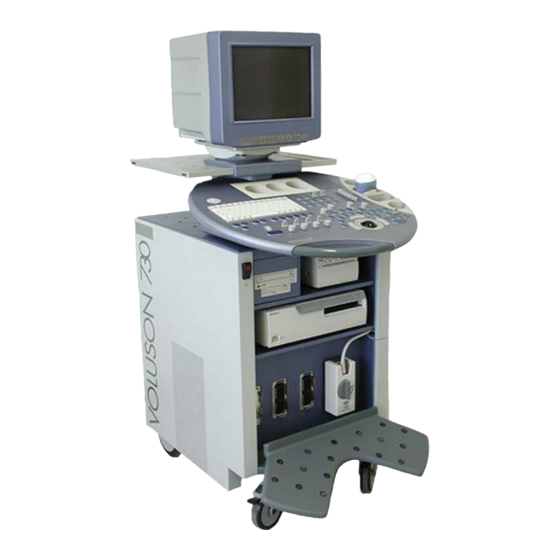
Description of the System System Assembly 3.3.1 Basic System It consists of the following modules: Probe connector module: This module houses the entire electronics for all connections, up to 3 probe connectors and the beamformer module (transmit and receive electronics). Control console: The control console consists of the hard keys, digipots, navigation wheel and the trackball, the loudspeakers, and the probe holders. -
Page 51: Optional Modules
Description of the System 3.3.3 Optional Modules Optional modules (CW Doppler, Real Time 4D, etc.) according to the pricelist of the Voluson 730Pro. 3.3.4 Position of Display Annotation ® Voluson 730Pro - Basic User Manual 105831 Rev. 0... -
Page 52: Concept Of Operation
Description of the System Concept of Operation The control center is the console with the hard keys, the digipots, the flip switches, the keyboard (with shortcut functions) and the trackball. The console controls frequently used functions, e.g., Freeze/Run, change of modes, etc. Additional functions are controlled via the navigation wheel. -
Page 53: Changing Of Menus
Description of the System 3.4.1 Changing of Menus Each menu has its own [Sub Menu]. By selecting this menu item the related menu appears in the menu area on the left side of the screen. Change Menu For example: If the current image mode is 2D/C/PW, this button changes the image menu sequentially into 2D-, Color-, and Doppler Image Menu. - Page 54 Description of the System Image Info: B-Mode User program Name of user program 7.5-5.0 Receiver frequency [MHz] Pwr -3 Acoustic power [dB] Gn -12 Gain [dB] Selected Dynamic curve [number] and Gray map [number] C6 / M1 P4 / E2 Persistence [number] and Edge enhancement [number] MI 0.4 Mechanical Index...
-
Page 55: Control Panel
Description of the System 3.4.3 Control Panel Loudspeaker position TGC slider controls Mode keys (digipot controls) Trackball Trackball Menu Navigation key Menu digipot and flip switch controls Navigation wheel (Menu control element) Keyboard with shortcut function keys Ultrasound scanning gel holder ®... -
Page 56: Hard Keys
Description of the System 3.4.4 Hard keys Read/Write (Freeze/Run) when bright: image is frozen (read mode) when dark: real time scan (write mode) review: To Freeze an Image (chapter 4.5.2) Printer Trigger A remote trigger key for B/W Printer, SCSI Color Printer, DICOM Printer key setup review: Peripherals (chapter 17.3.3) - Page 57 Description of the System Clear All to clear graphics, measurements and annotations on the screen Indicator display a pointer arrow or hand operation review: Indicator (chapter 4.7.3) Bodymark to enter Bodymark symbols on the screen operation review: Pictogram (chapter 4.7.4) ABC - Image Annotation write onto the screen for documentation operation review:...
- Page 58 Description of the System Automatic Optimization in 2D Mode: Pressing this key causes automatic optimization of the gray scale to enhance the contrast resolution. operation review: (chapter 5.6) in PW Mode: Press this key for automatic optimization of the PRF and Baseline. operation review: (chapter 7.2.4) Exit...
- Page 59 Description of the System 3D/4D Image Orientation press this key to change the image orientation of the 3D-image Up/Down Image Orientation to alternate between up and down image orientation Left/Right Image Orientation to alternate between left and right image orientation Initial Position resets the rotations and translations of a volume section to the initial position Beam steering...
- Page 60 Description of the System Microphone switch Microphone on/off No function switch ECG signal on/off Sonoview press this key to shift from scan mode to Sonoview operation review: Sonoview (chapter 15) Report patient report page operation review: OB Patient Report (chapter 14.4) GYN Patient Report (chapter 14.6) Cardiac Patient Report...
- Page 61 Description of the System Real Time 4D Mode activates the Real Time 4D mode (continuous volume sweep) operation review: Real Time 4D Acquisition (chapter 11.5) Power Doppler pressing activates the Power Doppler mode provided the active probe allows for it review: PD Mode (chapter 9)
-
Page 62: Function Of The Trackball At Diverse Dialog
Description of the System 3.4.4.1 Function of the Trackball at Diverse Dialog Pages In general operations at diverse dialog pages and windows on the system desktop (e.g., Patient Data Entry, Usage of EUM, System Setup, Measurement Setup, etc.) are done with the trackball and the trackball keys (mouse emulation). -
Page 63: Keyboard Keys
Description of the System 3.4.4.2 Keyboard keys Delete Line deletes the complete image annotation line Delete Image Annotation deletes all the image annotations on the screen Delete Arrow deletes the displayed pointer arrows Delete Measurement deletes the measurements on the screen Delete deletes all graphics, measurement, pointers and image annotations on the screen Press the [F1] button to invoke the electronic user manual. -
Page 64: Electronic User Manual (Eum)
Description of the System Electronic User Manual (EUM) Press the [F1] key on the keyboard to invoke the electronic user manual. The EUM screen appears (e.g.: 2D Mode). With the [Back] button it is possible to change to the previous selected topic. With the [Print] button it is possible to print out the selected topics on the default line printer. -
Page 65: Searching With The Help Topic: Contents
Description of the System 3.5.2 Searching with the Help Topic: Contents Each „Book“ includes the topics of the selected chapter. 1. Select the desired chapter using the trackball and the [Set] key (right/left trackball key). 2. Click the [Open] button to display the topics of the selected chapter. 3. -
Page 66: Searching With The Help Topic: Index
Description of the System 3.5.3 Searching with the Help Topic: Index 1. Type the first few letters of the word you’re looking for. 2. Click the desired index entry. 3. Click the [Display] button. The explanation of the selected topic appears on the screen. Voluson®... -
Page 67: Searching With The Help Topic: Find
Description of the System 3.5.4 Searching with the Help Topic: Find Field 1: Type the search word(s). Field 2: Select matching words to narrow search. Field 3: Click a topic. Click the [Display] button. The explanation of the selected topic appears on the screen. ®... -
Page 68: Setup Of The Find Options
Description of the System 3.5.4.1 Setup of the Find Options Select the [Options] button in the Help Topic “Find”. The menu of the available options for the Help Topic “Find” appears on the screen. Voluson® 730Pro - Basic User Manual 3-24 105831 Rev. - Page 69 Starting the System Starting the System .......................4-2 General Remarks ......................... 4-2 Safety Warnings........................4-2 Turn on Power ........................4-2 Transducer Connection....................... 4-3 Probe/Program Selection ....................4-4 4.5.1 Starting the System......................4-5 4.5.2 To Freeze an Image ......................4-6 Entering Patient Data......................4-6 4.6.1 Patient ID Menu ......................
-
Page 70: Starting The System
Starting the System Starting the System General Remarks Installation, switching on the first time and check-up of the system must only be performed by authorized persons. The VOLUSON® 730Pro is delivered with recommended basic settings. These offer suitable conditions for a large number of applications. Depending on the user's experience these default settings can be changed and stored as new User Programs. -
Page 71: Transducer Connection
Starting the System Transducer Connection CAUTION! Prior to connecting or disconnecting a probe freeze the image. It is unnecessary to switch the unit off. If a probe is disconnected while running (write mode) a software error may occur. In this case switch the unit OFF and later ON (perform a reset). CAUTION! If the cable spout on the right-hand door is missing do not pull on the probe cable, the probe cable can be damaged. -
Page 72: Probe/Program Selection
Starting the System Probe holders: Always store the probes in the intended probe holders. Probe/Program Selection This menu shows the connected probes. The denomination of each transducer connected to the system appears on the screen. Probe selection is done with the trackball. The field with the selected probe is illuminated. At the same time the available applications of the selected probe are displayed in the [Application] field. -
Page 73: Starting The System
Starting the System Probe selection menu on the screen: Probe window: Shows all connected probes, the active one is highlighted (if one is active). Application window: Shows all applications for the active probe. The last active application is marked. Setting (program) window: Shows all settings for the active application. -
Page 74: To Freeze An Image
Starting the System 4.5.2 To Freeze an Image Freeze/Run key (hard key) Storing of the image by pressing the [Freeze] key: Bright key: read mode (image is stored, probe deactivated) Dark key: write mode (real time is on, probe activated) Entering Patient Data Patient Data is to be entered by using a patient data form. - Page 75 Starting the System End Exam / New ID: Patient and measurement data are stored in the “Data manager” (all temporary patient and measurement data are cleared). Display: “Patient Information” screen End Exam / Exit: Patient and measurement data are stored in the “Data manager” (all temporary patient and measurement data are cleared).
-
Page 76: Edit Patient Info
Starting the System 4.6.1.1 Edit Patient Info Select the [Edit] item which is available in the Patient ID Menu. see: (chapter 4.6.1) Procedure see: Standard Input (chapter 4.6.2) Continue the actual Exam: The patient info is temporarily stored. The Edit Patient Info procedure is closed and the previous operating state is activated. Note: The ID number cannot be changed! ®... -
Page 77: Standard Input
Starting the System 4.6.2 Standard Input ♦ SELECT AN INPUT FIELD There are 2 possibilities to select an input field: 1. by means of the trackball 2. by means of the keyboard 1. Input field selection possibility: Trackball: position the cursor in the input field Enter: select an input field, press the right or left trackball key 2. - Page 78 Starting the System Note: Patient data on different systems such as the Voluson 730Pro or 3D View 2000 are only distinguished by the Patient Identification (ID) field! If you do not use the default auto-generated ID please make sure that this identification (ID) is...
-
Page 79: To Retrieve Patient Data Via External Worklist Server
Starting the System 4.6.3 To Retrieve Patient Data via External Worklist Server Select the [Worklist] button to view the available Data from the external worklist server. This button is available in the “Patient Information” screen. 1. Select the desired “Search” field with the trackball. 2. -
Page 80: Patient Information - General
Starting the System 4.6.3.1 Patient Information - General • Application: General Data Field ID number max. 32 characters Name patient name max. 32 characters Accession # accession number max. 16 characters Day of Birth patient day of birth patient age ---- , female, male (selection in pull down menu) Review: Standard Input... -
Page 81: Patient Information - Ob
Starting the System 4.6.3.2 Patient Information - OB • Application: Application Field: The date of the last menstrual period must be entered using format yyyy-mm-dd. Note: The first day of the last period must be entered. GA and EDD are calculated automatically after entering the LMP date. GA (LMP) Enter the gestational age (weeks and days), LMP and EDD is calculated automatically. -
Page 82: Patient Information - Gyn
Starting the System 4.6.3.3 Patient Information - GYN • Application: Application Field: The first day of the last menstrual period must be entered using format yyyy-mm-dd. Exp. Ovul. Date of expected ovulation. Day of cycles Day of cycles. Gravida Enter the patient's history of pregnancies. Para Enter the patient's history of live births. -
Page 83: Patient Information - Cardio
Starting the System 4.6.3.4 Patient Information - Cardio • Application: Cardio Application Field: Height Enter the patient’s height in one of the units (cm, ft, inch) Weight Enter the patient’s weight in one of the units (kg, lp, oz) Body Surface Area (calculation value, no input) Heart Rate Unit selection: Position the cursor into the unit field using the trackball and press the left or... -
Page 84: To Search In The Patient List
Starting the System 4.6.4 To Search in the Patient List Select the [Search] button using the trackball cursor and enter with the left or right trackball key. This button is available in the “Patient Information” screen. The “Search Results Dialog” menu appears on screen. Search procedure: •... -
Page 85: Image Annotation
Starting the System Image Annotation [ABC] - Annotation key (hard key) Press this key to start the documentation function. Upon pressing once more the text is switched off but the written text is not cleared. There are two possibilities provided to write on the screen: Annotation: with the keyboard keys Auto Annotation: with the navigation wheel, annotation with predefined words... -
Page 86: Auto Annotation
Starting the System 4.7.2 Auto Annotation This function is provided to rapidly insert terms into the display image. 40 words for each application are user-programmable. Programming of the TEXT AUTO function review: User Setting (chapter 17.3.2). Operation: Activate the Text Mode via [ABC] key. The auto text menu appears in the menu area. - Page 87 Starting the System With the corresponding flip switch you can select either 1 or 2 page of words. Text function is switched off but the entered text is not cleared. Return to the last active menu. Press either the [Delete ABC] key on the keyboard or the [Clear all] key on the control panel to delete all the entered text.
-
Page 88: Indicator
Starting the System 4.7.3 Indicator Indicator key (hard key) Pressing the [Indicator] key causes the menu area to change to the Indicator menu. The last used Indicator (or by default the first indicator on the touch menu) appears on the screen. Operation: Switch on the indicator function (hard key). -
Page 89: Pictogram
Starting the System 4.7.4 Pictogram For the documentation of the scan position on the patient, a selection of graphic body symbols (bodymark) is available. The scan position is indicated by a short bright line. This line can be positioned freely on the bodymark symbol. Pressing the [Bodymark] key causes the menu area to change to the bodymark menu. - Page 90 Starting the System After selecting the [New Symbol] item the application-related bodymarks appear on the screen. Return to the last active menu with the bodymark displayed. Return to the last menu with the bodymark switched off. Changes between the different application-related bodymarks. After selecting another application the menu changes back to the bodymark menu with the bodymark symbols of the selected application.
- Page 91 2D Mode 2D Mode ........................5-2 2D Main Menu ........................5-2 2D Mode Depth ........................5-4 2D Image Angle........................5-4 2D Gain..........................5-5 TGC Slider Controls......................5-5 2D Automatic Optimization....................5-6 Transmit Power ........................5-6 Transmitter Focus ....................... 5-7 Receiver Frequency Range ....................5-7 5.9.1 Harmonic Imaging......................
-
Page 92: D Mode
2D Mode 2D Mode The 2D display consists of the ultrasound image, an orientation marker, patient data, image information, a Gray scale pattern, a depth scale with focal zone markers and an actual TGC curve. The ultrasound image is derived from the tissue echoes that return to the scanhead. They are amplified, converted and then mapped to an image processing curve that relates each echo’s intensity to a shade of gray. - Page 93 2D Mode The 2D Main menu appears on the screen. (write mode) Remarks: In read mode changing the Angle, Beta View, Focal Zones, OTI, Frequency, Trapezoid mode and FFC (Focus and Frequency Composite) is impossible. The functions Focal Zones, OTI, Beta View, Angle, Frequency, FFC and Trapezoid mode only appear if they are available for the selected probe.
-
Page 94: Mode Depth
2D Mode 2D Mode Depth With this function the depth range of the ultrasound image for the region of interest is adjusted. The number of image lines and the frame rate are automatically optimized. Changing of the depth is only possible in real time (write mode). -
Page 95: Gain
2D Mode 2D Gain With the “Gain” control the overall brightness of the 2D image is adjusted. The adjustment of the gain control determines the amount of amplification applied to the received echoes. All incoming echoes are amplified with the same gain value regardless of depth. [2D Mode] key –... -
Page 96: Automatic Optimization
2D Mode 2D Automatic Optimization This function will optimize the contrast resolution according to the histogram of the scan area. The shape of the ROI will depend on the probe, scan depth and scan angle. The primary result is a value for the left and right endpoint of the actual histogram. Pressing the [auto] key causes automatic optimization of the gray scale to enhance the contrast resolution. -
Page 97: Transmitter Focus
2D Mode Transmitter Focus The selected focal zone determines the depth range of optimized sharpness of the ultrasound beam. The [Foc#] field in the status area displays the actually set number of focal zones for transducers which allow to change focal zones. Use the [Foc flip switch to select the number of focal zones. -
Page 98: Harmonic Imaging
2D Mode 5.9.1 Harmonic Imaging Tissue not only scatters back echoes with the nominal transmitted frequency but also with double, threefold, fourfold and so on (harmonic) frequencies, as a result of a physical effect called “non-linear propagation”. Harmonic Imaging delivers better grayscale contrast compared to standard ultrasound imaging. This technique has proved to be particularly useful for difficult-to-image patients and furthermore is less prone to artifacts. -
Page 99: Beta View (Β-View)
2D Mode 5.11 Beta View (β-View) The “Beta View” function allows the adjustment of the Volume O-Axis position of 3D probes in 2D mode. The green line in the displayed symbol indicates the position of the acoustic block. + and - defines the corresponding sweep direction on the screen. Rotate: changes the position of the acoustic block Press: moves the acoustic block back to 0°. -
Page 100: Trapezoid Mode
2D Mode 5.13 Trapezoid Mode Advantage of the Trapezoid mode: The scan area is increased in relation to the linear display by steering the ultrasound lines in the border of the probe. Selection of Linear- or Trapezoid mode display The actual mode is marked (e.g., Trapezoid). Remarks: This key appears automatically in the 2D mode menu, if the selected probe is capable of Trapezoid mode. -
Page 101: Multi Format
2D Mode Remarks: The Orientation marker is green in an active 2D image. The Orientation marker is white in a frozen Dual or Quad image. 5.15 Multi Format The "Multi Format" controls [Dual] and [Quad] allows you to display several 2D mode images simultaneously on the screen. - Page 102 2D Mode The upper trackball key: The upper trackball key changes between Cine and 2D image position of the actual 2D image. Freeze key: real time mode: The [Freeze] key freezes the real time 2D image in the actual display position. Freeze Mode: The [Freeze] key activates real time mode of the frozen 2D image in the actual display position.
-
Page 103: Quad-Screen Format
2D Mode 5.15.2 Quad-Screen Format [Quad] Screen Format key (hard key): Press this key to change from Single or Dual display mode to “Quad” mode. real time mode (write mode): The Quad mode key freezes the real time 2D image in the actual display position and shows the real time 2D image live in the next display position. - Page 104 2D Mode Operation: Select Quad display mode. Freeze the image. Select the next display position by using the format key. Freeze the next image. If the image is frozen and you use [Update 2D] (the right trackball key) the next image is selected and active.
-
Page 105: Cine Mode
2D Mode 5.16 Cine Mode While scanning a certain number of frames (2D images of the last examination sequence) will be stored in the cine memory upon [Freeze]. The sequence can be reviewed image by image. Move the trackball horizontally to display the 2D images of the stored sequence, one by one. -
Page 106: Auto Cine
2D Mode 5.16.2 2D Auto Cine With the “2D Auto Cine” function the user can review a defined sequence (start, end) of Single-, Dual- and Quad format 2D- and 2D/Color images. Review speed and read-zoom are available. 1. Store a 2D image or a CFM image. Note: In dual- and quad formats select the desired image using the [Format] keys. -
Page 107: Pan Zoom
2D Mode 9. Start/Stop the 2D Auto Cine function with the right or left trackball key. At dual- and quad formats only the Cine sequence of the active 2D image (indicated with the green dot) is displayed. Remarks: The 2D Auto Cine function is only possible in read mode. The 2D Auto Cine function is also possible at multiple format. -
Page 108: High Resolution Zoom
2D Mode 5.18 High Resolution Zoom The 2D image can be magnified in write mode. The displayed zoom box can be placed over the entire 2D image area, also the size of the zoom box can be changed. The scan frame rate and line number are automatically optimized with the zoom box active in write mode. -
Page 109: Sub Menu
2D Mode 5.19 2D Sub Menu The Main 2D mode must be active. Select the [Sub Menu] item. The functions of the 2D Sub menu are shown in the menu area as well as in the status area. Note: Changes are only possible in write mode (controls have no function in read mode). The following functions are available: Quality (chapter 5.19.1) -
Page 110: Line Filter
2D Mode 5.19.2 Line Filter With “Line Filter” especially the lateral resolution can be optimized with this innovative correlation algorithm. With the new process, the signals of the neighboring pulses are less weighted for the display of the actual pulse which considerably improves the detail resolution and signal-to-noise ratio. Three steps are provided: off, low, high The line filter state is displayed in the Image Info area on the screen. -
Page 111: Persistence Filter
2D Mode Remarks: The appearance of the gray values depends also on the selected gray map. To select a 2D gray map review: Gray Chroma Map (chapter 12.1) Changes are only possible in write mode. (Control has no function in read mode.) 5.19.5 Persistence Filter “Persistence”... - Page 112 2D Mode This page intentionally left blank. ® Voluson 730Pro - Basic User Manual 5-22 105831 Rev. 0...
- Page 113 M Mode M Mode .........................6-2 M Main Menu ........................6-2 6.1.1 Principle.......................... 6-3 M Operation......................... 6-4 6.2.1 Cursor Position ....................... 6-4 6.2.2 Activation of M Mode ....................6-4 6.2.3 Sweep Speed........................6-5 6.2.4 Invert ..........................6-5 6.2.5 Frequency ........................6-5 6.2.6 TGC Slide Controls ......................
-
Page 114: M Mode
M Mode M Mode M mode imaging provides Time and Motion echo information derived from a stationary ultrasound beam. M mode is used along with a 2D image. A straight line running through the 2D image, called the M-cursor, identifies the position of the stationary ultrasound beam from which the echo information is being gathered. -
Page 115: Principle
M Mode The “M Main” menu appears on the screen. (write mode) Remarks: In read mode changing the Speed and Frequency is not possible. 6.1.1 Principle The M mode display is derived from a 2D image display. When switching on the M mode, the M-cursor line is inserted into the 2D image. It symbolizes the ultrasound beam and defines the position of the M mode trace. -
Page 116: M Operation
M Mode M Operation The M operation consists of : Cursor Position (chapter 6.2.1) Activation of M Mode (chapter 6.2.2) Sweep Speed (chapter 6.2.3) Invert (chapter 6.2.4) Frequency (chapter 6.2.5) TGC Slide Controls (chapter 6.2.6) M Mode Depth (chapter 6.2.7) M Gain Control (chapter 6.2.8) M Cineloop... -
Page 117: Sweep Speed
M Mode 6.2.3 Sweep Speed On the status area you will find the [Speed] key. review: M Main Menu (chapter 6.1) By touching up or down four different sweep speeds can be selected. 1 = 3,5 cm/s 2 = 5,0 cm/s 3 = 7,5 cm/s 4 = 10,0 cm/s (in relation to the system’s monitor) -
Page 118: M Gain Control
M Mode 6.2.8 M Gain Control With the [Gain] control the overall brightness of the M mode trace can be adjusted. The adjustment of the Gain control determines the amount of amplification applied to the received echoes. All received echoes are amplified with the same gain value regardless of the scan depth. The M Gain function influences the M trace only. -
Page 119: M Sub Menu
M Mode M Sub Menu The M Main menu must be active. Select the [Sub Menu] item. The functions of the “M Sub” menu are shown in the menu area as well as in the status area. Note: Changes are only possible in write mode ( Controls have no function in read mode). Only changes in the Gray Chroma Map are possible in read mode. -
Page 120: Enhance
M Mode 6.3.2 Enhance With the “Enhance” function the echo information is digitally processed such that certain existing information becomes easily visible (e.g., adjacent media layers). Due to the Enhance function a finer, sharper impression of the image is produced. The Enhance state is displayed in the Image Info area on the screen. - Page 121 Spectral Doppler Mode Spectral Doppler Mode ....................7-2 PW Mode (Pulsed Wave Doppler) ..................7-2 7.1.1 PW Main Menu ......................7-3 7.1.2 PW Operation ......................... 7-4 7.1.2.1 Gate Position and Gate Width................. 7-4 7.1.2.2 Activation of PW Mode ..................7-5 7.1.2.3 PW Gain Control.....................
-
Page 122: Spectral Doppler Mode
Spectral Doppler Mode Spectral Doppler Mode Doppler imaging includes a spectral analysis which describes the Doppler shift signal from the moving reflectors within a sample volume. The spectral display scrolls from right to left and depicts the spectral distribution of the components of the Doppler shift frequency over time. Frequency or velocity values appear on the vertical axis and time along the horizontal axis. -
Page 123: Pw Main Menu
Spectral Doppler Mode 7.1.1 PW Main Menu Mode key (hard key) Press the [PW] control to activate the PW mode in the preparation mode. First only the PW-cursor appears on the active 2D image. To start and use the PW mode review: PW Operation (chapter 7.1.2) To adjust the PW settings review:... -
Page 124: Pw Operation
Spectral Doppler Mode 7.1.2 PW Operation The PW Operation includes: Gate Position & Gate Width (chapter 7.1.2.1) Activation of PW Mode (chapter 7.1.2.2) PW Gain Control (chapter 7.1.2.3) PW Automatic Optimization (chapter 7.1.2.4) Sweep Speed (chapter 7.1.2.5) Audio Signal (chapter 7.1.2.6) Invert (chapter 7.1.2.7) Angle Correction... -
Page 125: Activation Of Pw Mode
Spectral Doppler Mode 7.1.2.2 Activation of PW Mode By pressing the left or right trackball key the screen is divided asymmetrically. The 2D image appears above The PW spectrum appears below. Three display formats are possible; review: Format (chapter 7.1.3.2) The status area shows the PW mode activation controls. -
Page 126: Pw Automatic Optimization
Spectral Doppler Mode 7.1.2.4 PW Automatic Optimization This function will optimize the following settings: PRF: automatic detection of highest flow velocities and adjustment of the velocity scale Baseline: will be shifted so that the flow spectrum is centered Pressing the [auto] key enables automatic optimization of the PRF and Baseline. When the key is pressed again, the optimization will be updated. -
Page 127: Invert
Spectral Doppler Mode 7.1.2.7 Invert This function inverts the PW spectrum display in relation to the direction of the flow. The displayed spectrum is inverted around the baseline. The velocity or frequency scale changes accordingly. Use [Invert] when necessary to change the spectral display orientation. It is possible in both read- and write mode. -
Page 128: Baseline
Spectral Doppler Mode 7.1.2.9 Baseline The PW spectrum baseline shift enlarges the velocity range in one direction. The displayed velocities (cm/s, m/s) or frequencies (kHz) on the upper and lower edge of the screen (scale, white borderline) mark the maximum velocity (maximum measuring range) . The baseline can be shifted up in 8 steps or down in 8 steps. -
Page 129: Hprf Mode
Spectral Doppler Mode 7.1.2.11.1 HPRF Mode The maximum clearly measurable flow velocity (Nyquist Limit) is determined by the measuring depth of the sample volume and the related run time of the ultrasound. By a further increase of the Doppler PRF (High PRF mode, HPRF) the Nyquist Limit can be increased. -
Page 130: Freeze
Spectral Doppler Mode Important note: The determination of the envelope curve requires a clear and low-noise recording of the Doppler spectrum. Otherwise the reliability of the displayed measurement results may not be ensured! Remarks: Activating the Real Time Trace is only possible in write mode. No function in read mode. 7.1.2.13 Freeze The [Freeze] key starts and stops the 2D image and PW Doppler spectrum. -
Page 131: Pw Sub Menu
Spectral Doppler Mode 7.1.3 PW Sub Menu The PW Main menu must to be active. Select the [Sub Menu] item. The functions of the PW Submenu are shown in the menu area as well as in the status area. Note: Changes are only possible in write mode! However, changes of the angle and the baseline are also possible in read mode. -
Page 132: Format
Spectral Doppler Mode 7.1.3.2 Format This controls serve for selection of either one of three formats (60/40, 50/ 50 and 40/ 60) for display. Format: large (60/40) Format: mid (50/50) Format: small (40/60) 7.1.3.3 Frequency This control serves for selection of the required transmit frequency for the actual gate position. Normally one works with the transmit frequency which is corresponding to the ultrasound element’s properties [Frequency mid]. -
Page 133: Dynamic
Spectral Doppler Mode 7.1.3.4 Dynamic Dynamic refers to the compression of grayscale information into a suitable range for the display. Dynamic allows you to enhance a part of interest of the grayscale to make it easier to display pathology. It adjusts the displayed cutoff of the Doppler analysis waveform. Turn clockwise to decrease the brightness (more gray shades) ;... -
Page 134: Cw Mode (Continuous Wave Doppler)
Spectral Doppler Mode CW Mode (Continuous Wave Doppler) The CW mode is subdivided in two groups. In these groups you will see how to use CW mode and how to adjust the CW settings. To use the CW mode review: CW Main Menu (chapter 7.2.1) To adjust CW setting review:... -
Page 135: Cw Operation
Spectral Doppler Mode 7.2.2 CW Operation The CW operation includes: Cursor Position & Focus (chapter 7.2.2.1) Activation of CW Mode (chapter 7.2.2.2) CW Gain Control (chapter 7.2.2.3) Sweep Speed (chapter7.2.2.4) Audio Signal (chapter 7.2.2.5) Invert (chapter 7.2.2.6) Angle Correction (chapter 7.2.2.7) Baseline (chapter 7.2.2.8) (chapter 7.2.2.9) -
Page 136: Cw Gain Control
Spectral Doppler Mode 7.2.2.3 CW Gain Control CW Gain controls the amplification of the incoming Doppler signals. The Doppler gain should be adjusted to a level that fills in the grayscale of the spectral analysis waveform without creating a noise. -Mode] key adjusts the CW Gain. -
Page 137: Invert
Spectral Doppler Mode 7.2.2.6 Invert This function inverts the CW spectrum display in relation to the direction of the flow. The displayed spectrum is inverted around the baseline. The velocity or frequency scale changes accordingly. Use [Invert] when necessary to change the spectral display orientation. It is possible in both read- and write mode. -
Page 138: Baseline
Spectral Doppler Mode 7.2.2.8 Baseline The CW spectrum baseline shift enlarges the velocity range in one direction. The displayed velocities (cm/s, m/s) or frequencies (kHz) on the upper and lower edge of the screen (scale, white borderline) mark the maximum velocity (maximum measuring range). The baseline can be shifted up in 8 steps or down in 8 steps. -
Page 139: Real Time Trace
Spectral Doppler Mode 7.2.2.11 Real Time Trace With the “Real Time Auto Trace” function the envelope curve of the Doppler spectrum (maximum velocities) and the corresponding evaluation is automatically displayed on the monitor. Select the [RT Trace] item to display the curve for the maximum velocities (envelope curve) simultaneously with the Doppler spectrum. -
Page 140: Cw Sub Menu
Spectral Doppler Mode 7.2.3 CW Sub Menu The “CW Main” menu must be active. Select the [Sub Menu] item. The functions of the CW Sub menu are shown in the menu area as well as in the status area. Note: Changes are only possible in write mode ! However, changes of the angle and the baseline are also possible in read mode. -
Page 141: Format
Spectral Doppler Mode 7.2.3.2 Format This controls serve for selection of either one of three formats (60/40 , 50/ 50 and 40/ 60) for display. Format: large (60/40) Format: mid (50/50) Format: small (40/60) 7.2.3.3 Dynamic Range Dynamic refers to the compression of grayscale information into a suitable range for the display. Dynamic allows you to enhance a part of interest of the grayscale to make it easier to display pathology. -
Page 142: Exit The Cw Submenu
Spectral Doppler Mode 7.2.3.4 Exit the CW Submenu Press the [Exit] key on the control panel to exit the CW Submenu. 7.2.4 CW + 2D + Color Information (Triplex Mode) Triplex mode is the simultaneous real-time display of 2D mode, Spectral Doppler and Color Doppler. There are two possibilities to combine Continuous Wave Doppler (CW) with Color Information: •... - Page 143 CFM Mode CFM Mode (Color Flow Mode) ..................8-2 CFM Main Menu ......................... 8-2 CFM Operation........................8-3 8.2.1 CFM Box Position and CFM Box Size ................8-4 8.2.2 CFM Gain Control......................8-4 8.2.3 Quality ..........................8-5 8.2.4 WMF ..........................8-5 8.2.5 Velocity Range (PRF) ....................
-
Page 144: Cfm Mode (Color Flow Mode)
CFM Mode CFM Mode (Color Flow Mode) Color imaging uses the Doppler principle to build a Color image. The Color coding gives information about blood flow velocity, direction, quality, and timing. This information is then used to overlay a Color image onto the 2D grayscale scan image. Color imaging helps you to locate blood flow disturbances. -
Page 145: Cfm Operation
CFM Mode The “CFM Main” menu appears on the screen (write mode). Remarks: Changing the Quality, Frequency, Wall Motion Filter, PRF, Gain, Invert and 2D+2D/C is only possible in write mode. Beam steering is only possible with linear probe and in write mode. CFM Operation The CFM operation consists of : CFM Box Position and CFM Box Size... -
Page 146: Cfm Box Position And Cfm Box Size
CFM Mode 8.2.1 CFM Box Position and CFM Box Size In 2D imaging the relationship between 2D frame rate, line density, and field-of-view are well known factors to be considered to obtain optimum 2D images. Similar relationships exist in Color imaging. On the CFM Submenu the selection of the line density adjusts the balance between the 2D Line Density and the Color mode line density. -
Page 147: Quality
CFM Mode 8.2.3 Quality This control improves the Color Resolution by reducing the image frame rate alternatively it reduces the Color Resolution by increasing the image frame rate. Quality control (flip switch) There are three steps for the Color Quality: high: higher Color resolution/ lower frame rate norm:... -
Page 148: Velocity Range (Prf)
CFM Mode 8.2.5 Velocity Range (PRF) The velocity range of the display is governed by the pulse repetition frequency (PRF). The [PRF] control changes the display range. As you increase the velocity range by that control, the PRF increases. As the display scale increases, the maximum Doppler shift information that can be displayed without aliasing also increases. -
Page 149: Invert
CFM Mode 8.2.7 Invert This function inverts the color display in relation to the direction of the flow. The color of the color wedge inverts around the baseline. key unlit Normal: Flow to the transducer RED. Flow away from the transducer BLUE. key lit Inverted: Flow to the transducer BLUE. -
Page 150: Cfm Sub Menu
CFM Mode CFM Sub Menu “Main CFM” menu has to be active. Select the [Sub Menu] item. The functions of the CFM Submenu are shown in the menu area as well as in the status area. Note: Changes are only possible in write mode ! Changes in the Display Mode, Scale, CFM Map and Baseline are also possible in read mode. -
Page 151: Display Modes
CFM Mode 8.3.1 Display Modes The following Color display modes can be selected: Velocity, Turbulence (also Variance) and their combinations Velocity and Turbulence, Velocity and Power, Power and Turbulence. The Velocity display allows to see speed and direction of the blood flow. The Turbulence display allows to see the variation of the blood flow (turbulent flows). - Page 152 CFM Mode Display of Turbulence (T) The Turbulence is coded in a single color wedge: Less turbulent: dark green more turbulent: light green (low brightness) (high brightness) Display of Velocity and Turbulence (V-T) This is the Mode for high flow with turbulence (display of variance). low flow <...
- Page 153 CFM Mode Display of Velocity and Power (V-Pow) Direction and velocity are coded in two color wedges: low flow < ---- high flow > CFM Map 1 Forward flow: dark red – light red - Power: violet – light violet (pink) Reverse flow: dark blue –...
-
Page 154: Scale
CFM Mode 8.3.2 Scale The maximum velocities are displayed above and under the color scale. (kHz, cm/s, m/s) Select the scale display as desired. kHz: Doppler shift frequency cm/s: Flow velocity m/s: Flow velocity 8.3.3 Line Filter Especially lateral resolution can be optimized with this innovative correlation algorithm. With this new process, the signals of the neighboring pulses are less weighted for the display of the actual pulse which considerably improves the detail resolution and signal-to-noise ratio. -
Page 155: Baseline
CFM Mode 8.3.6 Baseline The CFM baseline shift can be used to prevent aliasing in one flow direction similar to the PW Doppler baseline shift. Shifting the CFM baseline enlarges the velocity range in one direction. The zero line of the color bar is also shifted. There are 8 Steps in each direction. -
Page 156: Cfm Map
CFM Mode 8.3.9 CFM Map This function provides selectability of the color coding for an optimization of the display of blood flow (similar to the post-processing curves with grayscale 2D). It is useful especially with low flow rates. It may be altered in real time or Freeze mode, respectively. Velocity display (Display V), velocity-turbulence display (Display V-T) and velocity-power display (Display V-Pow) each have different color pattern maps available for selection. -
Page 157: Dynamic Set
CFM Mode 8.3.11 Dynamic Set “Dynamic Set” controls the axial resolution of color in the display. It adjusts the axial sample depth of color pixels. When rotating counter-clockwise the color information is sharper (edges more pixely). When rotation clockwise the color information is less sharp (edges smoothed). Rotate the [Dyn.Set] digipot and select the desired dynamic range. -
Page 158: Cfm + 2D + Spectral Doppler (Triplex Mode)
CFM Mode 8.3.14 CFM + 2D + Spectral Doppler (Triplex Mode) Triplex mode is the simultaneous real time display of 2D mode, Color-Doppler and Spectral Doppler. There are two possibilities to combine Color Flow mode (CFM) with Spectral Doppler Information: •... - Page 159 PD Mode PD Mode (Power-Doppler Mode) ................9-2 PD Main Menu........................9-3 PD Operation ........................9-4 9.2.1 PD Box Position and PD Box Size................. 9-4 9.2.2 PD Gain Control ......................9-5 9.2.3 Quality ..........................9-5 9.2.4 Frequency ........................9-6 9.2.5 WMF ..........................9-6 9.2.6 Velocity Range (PRF) ....................
-
Page 160: Pd Mode (Power-Doppler Mode)
PD Mode PD Mode (Power-Doppler Mode) Sonographic diagnostics can be greatly extended by Color-Doppler sonography. Still, Color-Doppler sonography has deficits, especially in displaying very slow flow velocities - such as neovascularization to be found in malignant tumors. Power-Doppler intends to erase this deficit by displaying such slow flow velocities. In gynecologic and obstetric applications the advantages can be clearly seen in the display of placental blood circulation. -
Page 161: Pd Main Menu
PD Mode PD Main Menu PD Mode key (hard key) By pressing the control [PD] the PD Mode is switched on and the PD Box appears on the active 2D image. To use the PD mode review: PD Operation (chapter 9.2) To adjust the PD settings review: PD Sub Menu (chapter 9.3) -
Page 162: Pd Operation
PD Mode PD Operation The PD operation consists of : PD Box Position and PD Box Size (chapter 9.2.1) PD Gain Control (chapter 9.2.2) Quality (chapter 9.2.3) Frequency (chapter 9.2.4) (chapter 9.2.5) Velocity Range (PRF) (chapter 9.2.6) 2D + 2D/PD (chapter 9.2.7) Threshold (chapter 9.2.8) -
Page 163: Pd Gain Control
PD Mode 9.2.2 PD Gain Control PD Gain must be adjusted properly to ensure that continuous flow is displayed, where appropriate. PD Gain should be set as high as possible without displaying random Color speckle. If you set the PD Gain control to low, the lack of sensitivity will make it difficult to detect small abnormalities in flow and will result in an underestimation of the large flow disturbances. -
Page 164: Frequency
PD Mode 9.2.4 Frequency The selection of the transmit frequency depends also on the PD-Box position. It is common to use the transmit frequency which is the center frequency [Frequency mid] of the ultrasound crystal. With a higher transmit frequency [Frequency high] the amplitude of the Doppler spectrum is displayed larger (advantage: better display of lower flow velocities), but the penetration depth is reduced. -
Page 165: Velocity Range (Prf)
PD Mode 9.2.6 Velocity Range (PRF) The velocity range of the display is governed by the pulse repetition frequency (PRF). The [PRF] control changes the display range. Increasing the velocity range setting will increase the PRF. As the display scale increases, the maximum Doppler shift information that can be displayed without alaising also increases. -
Page 166: Pd Sub Menu
PD Mode PD Sub Menu The “PD Main” menu has to be active. Select the [Sub Menu] item. The functions of the PD Submenu are shown in the menu area as well as in the status area. Note: Changes are only possible in write mode. Only changes in the PD Map are possible in read mode. -
Page 167: Line Filter
PD Mode 9.3.1 Line Filter Especially lateral resolution can be optimized with this innovative correlation algorithm. With this new process, the signals of the neighboring pulses are less weighted for the display of the actual pulse which considerably improves the detail resolution and signal-to-noise ratio. Eight steps are provided. -
Page 168: Ensemble
PD Mode 9.3.4 Ensemble This function controls the number of pulses for one displayed Power-Doppler line. Since several pulses are to be evaluated for displaying a result, the color display quality increases with the number of evaluated pulses. Switch the [Ensemble] key and select the number of pulses per color line. max. -
Page 169: Balance
PD Mode The color is coded in a color wedge: PD Map1 PD Map 2 PD Map 3 PD Map 4 cyclamen gray-green brown dark red violet orange pink orange light red yellow light yellow yellow yellow PD Map 5 PD Map 6 PD Map 7 PD Map 8... -
Page 170: Dynamic Set
PD Mode 9.3.8 Dynamic Set “Dynamic Set” controls the axial resolution of color in the display. It adjusts the axial sample depth of color pixels. When rotating counter-clockwise the color information is sharper (edges more pixely). When rotation clockwise the color information is less sharp (edges smoothed). Rotate the [Dyn.Set] digipot and select the desired dynamic range. -
Page 171: Pd + 2D + Spectral Doppler (Triplex Mode)
PD Mode 9.3.11 PD + 2D + Spectral Doppler (Triplex Mode) Triplex mode is the simultaneous real time display of 2D mode, Power-Doppler and Spectral Doppler. There are two possibilities to combine Power-Doppler (PD) with Spectral Doppler Information: • PD + 2D Mode + PW Doppler (Pulsed Wave Doppler) •... - Page 172 PD Mode This page intentionally left blank. Voluson® 730Pro - Basic User Manual 9-14 105831 Rev. 0...
- Page 173 TD Mode TD Mode (Tissue Doppler Mode) ................10-2 10.1 TD Main Menu ....................... 10-2 10.2 TD Operation........................10-3 10.2.1 TD Box Position and TD Box Size ................10-4 10.2.2 TD Gain Control......................10-4 10.2.3 Quality .......................... 10-5 10.2.4 Velocity Range (PRF) ....................10-5 10.2.5 Invert ..........................
-
Page 174: Td Mode (Tissue Doppler Mode)
TD Mode TD Mode (Tissue Doppler Mode) Tissue-Doppler imaging generates a Color image by using the Doppler principle. This Color image is overlaid onto the 2D image. The Tissue image provides information about tissue motion direction and velocity. The Tissue-Doppler captures low flow but high amplitude signals associated with wall motion and creates a color-coded tissue image. -
Page 175: Td Operation
TD Mode The “TD Main” menu appears on the screen. (write mode) Remarks: Changing the Quality, PRF, Gain, Invert and 2D +2D/TD is only possible in write mode. 10.2 TD Operation The TD operation consists of : TD Box Position and TD Box Size (chapter 10.2.1) TD Gain Control (chapter 10.2.2) -
Page 176: Td Box Position And Td Box Size
TD Mode 10.2.1 TD Box Position and TD Box Size In 2D Imaging the relationship between 2D frame rate, line density, and field-of-view are well known factors to be considered to obtain optimum 2D images. Similar relationships exist in Color Imaging. On the TD Submenu the selection of the line density adjusts the balance between the 2D-Line Density and the Tissue mode line density. -
Page 177: Quality
TD Mode 10.2.3 Quality This control improves the Color Resolution by reducing the image frame rate alternatively it reduces the Color resolution by increasing the image frame rate. [Quality] control (flip switch) There are three steps for the Color Quality: high: higher Color resolution/ lower frame rate norm: normal Color resolution/ medium frame rate... -
Page 178: 2D/Td
TD Mode 10.2.6 2D + 2D/TD The “2D+2D/TD” function changes the single image display to two simultaneous half frames. The left frame shows only the 2D mode image. The right frame shows the 2D mode image also with color information. Switch on/ off this mode by touching the [2D+2D/TD] key. -
Page 179: Td Sub Menu
TD Mode 10.3 TD Sub Menu The “TD Main” menu must be active. Select the [Sub Menu] item The functions of the TD Submenu are shown in the menu area as well as in the status area. Note: Changes are only possible in write mode ! Only changes in the Gray Chroma Map, Scale, TD Map and Baseline are also possible in read mode. -
Page 180: Scale
TD Mode 10.3.1 Scale The maximum velocities are displayed above and under the color scale in (kHz, cm/s, m/s). Select the scale display as desired. kHz: Doppler shift frequency cm/s: Flow velocity m/s: Flow velocity 10.3.2 Line Filter Especially lateral resolution can be optimized with this innovative correlation algorithm. With this new process, the signals of the neighboring pulses are less weighted for the display of the actual pulse which considerably improves the detail resolution and signal-to-noise ratio. -
Page 181: Baseline
TD Mode 10.3.4 Baseline The TD baseline shift can be used to prevent aliasing in one flow direction similar to the Doppler baseline shift. Shifting the TD baseline enlarges the velocity range in one direction. The zero line of the color bar is also shifted. Adjust the zero line +/- There are 8 steps in each direction. -
Page 182: Line Density
TD Mode 10.3.6 Line Density This function determines the line density within the TD box. The lower the line density, the greater the distance between lines and the size of the color pixels. Switch the [Line D.] key and adjust the line density. max. -
Page 183: Balance
TD Mode 10.3.8 Balance “Balance” controls the amount of Color displayed over bright echoes and helps confine Color within the vessel walls. Decreasing this Balance displays Color on brighter structures. If you see no color, the Balance is probably set too high. Rotate the [Balance] digipot and select the useful Balance. -
Page 184: Smoothing
TD Mode 10.3.10 Smoothing A temporal average is determined from several color images, so that different filter periods can be selected for rising velocity and falling velocity. Rotate the [Smooth] digipots and select the rise and the fall filter. RISE: Filtering of the rise velocity leads to noise suppression. Avoid quick movements of the probe, because the motion is "built up"... - Page 185 Volume Mode Volume Mode......................11-3 11.1 Volume Acquisition with Volume Probes................ 11-4 11.1.1 Principle of Volume Acquisition.................. 11-5 11.1.2 Principal Scanning Modes.................... 11-5 11.1.3 What is Interactive 3D Image Rendering ? ..............11-6 11.1.3.1 What is Interactive ?....................11-6 11.1.4 Image Orientation (All Acquisition Modes)..............11-7 11.1.5 The Render Box......................
- Page 186 Volume Mode 11.5 Real Time 4D Acquisition....................11-52 11.5.1 Possible Display Adjustment Before Starting Acquisition.........11-54 11.5.2 Real Time 4D Acquisition During Active High Resolution Zoom ......11-55 11.5.2.1 Display of sectional Planes..................11-56 11.5.2.2 Display of REF-Image..................11-57 11.5.2.3 Display of ROI 4D....................11-58 11.5.2.4 Display of 4D ......................11-59 11.5.2.5 Display of A-ROI 4D ..................11-60...
-
Page 187: Volume Mode
Volume Mode Volume Mode General Description The Volume Mode allows for scanning a tissue volume and subsequent analysis of sections of the volume in 3 dimensions. The liberal selection of sections within the volume and the simultaneous real-time 4D display of three orthogonal planes and a rendered 3D Image represents a new dimension for e.g., the diagnosis of fetal abnormalities. -
Page 188: Volume Acquisition With Volume Probes
Volume Mode 11.1 Volume Acquisition with Volume Probes Volume mode keys (hard keys) By pressing on the [3D] respectively [4D] controls the Volume mode function is switched on and the Volume box appears on the Image area. The “3D Pre” or “4D Pre” menu appears on the screen (write mode). The Volume box appears on the image area. -
Page 189: Principle Of Volume Acquisition
Volume Mode 11.1.1 Principle of Volume Acquisition The acquisition of volume data sets is performed by 2D scans with special transducers designed for the 2D scans, the 3D sweep, and the real time 4D scans. The Volume acquisition is started using a 2D image with superimposed VOL-BOX. The 2D start image represents the central 2D scan of the volume. -
Page 190: What Is Interactive 3D Image Rendering
Volume Mode 11.1.3 What is Interactive 3D Image Rendering ? The 3D Image Rendering is a calculation process to visualize certain 3D structures of a scanned volume by means of a 2D image. The gray value for each pixel of the 2D image is calculated from the voxels along the corresponding projection path (analyzing beam) through the volume. -
Page 191: Image Orientation (All Acquisition Modes)
Volume Mode 11.1.4 Image Orientation (All Acquisition Modes) Start condition: B image: Adjust a longitudinal scan of the object desired. Switch on 3D mode and start the volume acquisition. B image orientation: up -> down Resulting orientation of sectional planes (read mode). ®... - Page 192 Volume Mode B image orientation: down -> up Resulting orientation of sectional planes (read mode). ® Voluson 730Pro - Basic User Manual 11-8 105831 Rev. 0...
-
Page 193: The Render Box
Volume Mode 11.1.5 The Render Box To obtain a good 3D picture, the following three points are very important (similar to a photography): • the direction of view • the area/size of view • free sight to the object (surface mode) This has to be adjusted with the render box. -
Page 194: General Advices To Obtain Good Rendered 3D Images
Volume Mode 11.1.6 General Advices to Obtain Good Rendered 3D Images B-MODE Poor quality of the volume scan will lead to a poor quality 3D image. For a good 3D image quality, adjust high contrast in 2D mode of the interesting structures before starting the volume scan. -
Page 195: Examples Of Rendered Images
Volume Mode 11.1.6.1 Examples of Rendered Images Surface mode: gray rendering Fetal hand Fetal face and umbilical cord Transparent mode: gray rendering Maximum mode: fetal skeleton Minimum mode: liver vessels ® Voluson 730Pro - Basic User Manual 11-11 105831 Rev. 0... -
Page 196: Volume Acquisition: 3D Sectional Planes
Volume Mode 11.2 Volume Acquisition: 3D Sectional Planes 1. Activate the Volume mode (hard key). 2. Select [Sect. Planes] Visualization mode from the menu area. 3. Select a 3D User Program (e.g., Default). The preset values are loaded. 4. Select the display format desired. Note: The selected format will be present in read mode after the acquisition is done. - Page 197 Volume Mode 8. Select the Quality (low, mid1, mid2, high1, high2). This function changes the line density against acquisition speed. Low: Fast speed / low scan density This mode is selected only in case of expected movement artifacts. A loss of volume resolution will result Mid2: Standard VOL scan / medium scan density High1:...
-
Page 198: Acquisition During Active High Resolution Zoom
Volume Mode 11.2.1 3D Acquisition During Active High Resolution Zoom 1. Press the [HR-Zoom] key. 2. Place the zoom box over the region of interest. The trackball has two functions: adjusting position or size of the zoom box. The activated function is displayed in the status area of the trackball. Press the upper trackball key to change the function of the trackball from position to size or vice versa. -
Page 199: After The 3D Sectional Planes Acquisition
Volume Mode 11.2.2 After the 3D Sectional Planes Acquisition After the 3D Sectional Planes acquisition, the system switches automatically to the “3D/4D Main” menu. The selected format will be present on the monitor (e.g., A,B,C - Sectional Plane Mode). Note: If you want to return to the “3D Pre” menu” press the right trackball key (Vol pre displayed in the status area of the trackball). -
Page 200: Orientation Help Graphic
Volume Mode 11.2.2.1 Orientation Help Graphic The orientation help image is only displayed in the lower right quadrant in Sectional Planes Mode. To activate or deactivate the Orientation Help Graphic select the [O.H. Graphic] item in the “3D/4D Main” menu. The volume body is shown in a somewhat simplified manner (arcs substituted by straight lines). - Page 201 Volume Mode Start situation Rotation (about X-axis) The position of the volume body in relation to the display plane is determined by a relative coordinate system. This is made up of three orthogonal axes, the common intersection of which is the 3-axial center of rotation.
-
Page 202: Image Position
Volume Mode 11.2.3.1 Image Position By this function the position of a reference image A, B or C in relation to the display field is determined. Press the upper trackball key to change the function from axis position to image position. -
Page 203: Selecting A Sectional Plane
Volume Mode 11.2.4 Selecting a Sectional Plane For the adjustment of an arbitrary sectional plane independent control functions are available. For faster rotating press on the digipot controls (toggle switch: slow rotation , fast rotation) Press again to return to slower rotation. 1. - Page 204 Volume Mode Note: For faster rotating press on the digipots control (switch function: slow rotation , fast rotation). Rotation about the X-axis of a reference image (e.g. A). Clockwise turn of the [X-axis] rotary control : By the clockwise rotation of the volume body in relation to the screen plane (as shown) the new sectional planes are calculated in real time and displayed on screen.
- Page 205 Volume Mode Rotation about the Z-axis of the reference image (e.g. A). Clockwise turn of the [Z-axis] rotary control : By the clockwise rotation of the volume body in relation to the screen plane (as shown) the new sectional planes are calculated in real time and displayed on screen. Important notes for the user: •...
-
Page 206: Translations
Volume Mode 11.2.6 Translations The translation allow a displacement of the center of rotation along the intersection lines of the sectional planes A, B and C. The displacement of the center of rotation leads to the display of parallel sectional images. To perform parallel slicing of images rotate the [Parallel shift] rotary control. -
Page 207: Initial Condition
Volume Mode I mportant note: The terms "front, left, top" etc., are not orientated to the patient, but serve for explanation. Of course, the "patient" could possibly be rotated to achieve the position described. By parallel movement of the reference image the new intersection lines will be displayed with the non- reference images. - Page 208 Volume Mode e.g., Init condition of an abdominal probe Screen display The sectional image A represents that 2D image, from which the volume acquisition was begun. If the VOL-start image is a longitudinal section (Cr on the left of the screen below), the following Init positions are obtained: anterior (ventral) posterior (dorsal)
-
Page 209: A,B,C - Sectional Plane Mode
Volume Mode 11.2.6.2 A,B,C - Sectional Plane Mode This display mode is active if you press the [Quad] screen format key. The 3 sectional planes A, B and C are standing normal on each other. The intersecting lines of the planes are the axes of the relative coordinate system and they are displayed in colors in the different images planes. - Page 210 Volume Mode 5. Select the reference image A , B or C by pressing the left trackball key repeatedly. (e.g. A) The selected reference image is surrounded with a green line. 3. Press the [Trackball Menu Navigation] key. Note: This key may have to be reselected depending on the function of the trackball. 4.
- Page 211 Volume Mode 6. To exit or to change the axis and position of the image within the Niche display, press the [Trackball Menu Navigation] key again. Position the images in the niche mode display with the trackball. Use the upper trackball key to change the function of the trackball from image position to axis position.
-
Page 212: Sub Menus
Volume Mode 11.3 Sub Menus The [Sub Menu] item is available in all Volume acquisition menus (read- and write mode). Select the [Sub Menu] item. The “3D/4D Sub” menu appears in the menu area as well as in the status area. Note: The Render Direction items are not available in Static 3D Sectional Planes mode. -
Page 213: Render View Direction
Volume Mode 11.3.1 Render View Direction The 3D render box determines the ROI (region of interest) for the 3D calculation and determines the direction of the view through the volume block. The adjustment of the render box is done with help of the 3 orthogonal planes A, B and C, each dividing the box in the middle. -
Page 214: Contrast
Volume Mode 11.3.3 Contrast Graphic display on screen: This graphic is provided only for the 3D/4D image. Horizontal axis: gray values from 0 to 255 Vertical axis : brightness from black to white 1. Set the start point of gamma curve bending. 2. -
Page 215: Volume Acquisition: 3D Rendering
Volume Mode 11.4 Volume Acquisition: 3D Rendering 1. Activate the Volume mode (hard key). 2. Select [3D Rendering] Visualization mode from the menu area. 3. Select a 3D User Program (e.g.: Default). The preset values are loaded. 4. Select the display format desired. Note: The selected format will be present in read mode after the acquisition is done. - Page 216 Volume Mode 8. Select the Quality (low, mid1, mid2, high1, high2). This function changes the line density against acquisition speed. Low: Fast speed / low scan density This mode is selected only in case of expected movement artifacts. A loss of volume resolution will result Mid2: Standard VOL scan / medium scan density High1:...
-
Page 217: After The 3D Rendering Acquisition
Volume Mode 11.4.1 After the 3D Rendering Acquisition After the 3D acquisition, the system switches automatically to the “Static 3D” menu. The selected format will be present on the monitor (e.g.: 3D ROI Mode). Note: If you want to return to the “3D Pre” menu press the right trackball key (Volpre displayed in the status area of the trackball). -
Page 218: Adjust Size And Content Of The Render Box
Volume Mode 3D Full-size Display: [Single] screen format key illuminated. The rendered 3D image is magnified and displayed in a full-size format without the sectional planes A, B, and C. 11.4.1.2 Adjust Size and Content of the Render Box Select reference image A, B or C with the left trackball key. The rotary controls and the trackball are assigned to the reference image for the adjustment of the render box functions. -
Page 219: Pictogram Mode
Volume Mode 11.4.1.3 3D Pictogram Mode Condition: A 3D-image being useful for a pictogram has to be displayed on screen, otherwise adjust a useful 3D image before. The [Accept ROI] item - in the “Static 3D” menu - and the [Quad] screen format key are selected. - Page 220 Volume Mode Adjust the magnification of the rendered 3D image The 3D image as well as the sectional image can be varied by their aspect ratio. Select a reference image A, B or C: The selected reference image is sectional plane A: The spatial position of plane A in relationship to the displayed 3D image is always vertical and also normal to the 3D image display.
- Page 221 Volume Mode Adjust the position of image B and C with trackball: The position of image B and C in relationship to the reference image A is determined through the Y-axis (= intersection line for image B) and through the X-axis (= intersection line for image C).
- Page 222 Volume Mode Selected reference image is sectional plane C: The spatial position of plane C in relationship to the displayed 3D image is always a parallel plane with a rotation of 90°. Therefore it is not possible to indicate the trace of image C by means of an intersectional line within the 3D image.
-
Page 223: Render Parameter - Basic Mode And Render Algorithm
Volume Mode 11.4.2 Render Parameter – Basic Mode and Render Algorithm For a good 3D image please note the following application hints: 1. Surface Modes: Adjust the render box in a way to have free sight from the render-start-area to the surface to be displayed. -
Page 224: Gray Render Mode
Volume Mode 11.4.2.1 Gray Render Mode In Gray Rendering Mode only the gray information of the data set is used, even if a Color Volume image is displayed. In case of a data set without color information, this mode is automatically activated. 1. -
Page 225: Threshold Control In Gray Render Mode
Volume Mode X-Ray Mode: Representation of all gray values within the ROI. Application: Tissue block with tumor or similar. The software module allows selection of 2 modes which are simultaneously calculated. Always the actual selected mode is displayed with 100%. With the [Mix]-key one can mix between the chosen modes. -
Page 226: Color Render Mode
Volume Mode 11.4.2.2 Color Render Mode In Color Rendering color information of Color or Power-Doppler signal can be used for the 3D display. 1. Activate Color Rendering Mode (if not active). 2. Select desired render algorithm. Note: Always select 2 modes! Surface Mode: Surface display of color information of blood flows. -
Page 227: Threshold Control In Color Render Mode
Volume Mode 11.4.2.2.1 Threshold Control in Color Render Mode Threshold Low (Reject): Normally this threshold must always be adjusted for a good appearance of the 3D surface image. By changing the [PD Low], all echoes below the threshold level are enhanced in pink color for a certain interval. -
Page 228: Glass Body Render Mode
Volume Mode 11.4.2.3 Glass Body Render Mode In Glass Body Render Mode the color and the gray information are processed into a 3D/PD or 3D/CFM volume. 1. Activate Glass Body Render Mode (if not active). 2. Select desired render algorithm. Note: Always select 2 modes! The following Rendering combinations are possible: Color acquisition in Power mode... -
Page 229: Rotation Cine
Volume Mode 11.4.3 3D Rotation Cine To get an overall 3D impression of the rendered object a certain number of calculated views are displayed image by image in a fast sequence. The rendered object, so to speak, is rotating for the observer. - Page 230 Volume Mode 3. Select the Rotation Angle Select the rotation angle you want. Alternatively use the [Start] and [End] digipot controls to select the rotation angle desired. 4. Select the Step Angle The step raster defines the amount of rotation between adjoining 3D images.
-
Page 231: Start/Stop The Calculated Sequence
Volume Mode 11.4.3.2 Start/Stop the Calculated Sequence Toggle function: [Start / Stop] Press the right or left trackball key to initialize the 3D cine sequence. On screen the cine sequence begins rotating ( if not rotating already). Loop Mode Direction If several 3D images have been calculated, they can automatically be displayed in a sequence. -
Page 232: Magicut
Volume Mode 11.4.4 MagiCut Principle This software has the ability to electronically edit the images, and makes it possible to cut away "3D artifacts". The left image displayed above is rendered without cutting, whereas the right image has had cutting techniques applied to give a clearer view of the object of interest. -
Page 233: Magicut Operation
Volume Mode 11.4.4.1 MagiCut Operation 1. Select the [MagiCut] item. The “MagiCut” menu appears in the menu area. Remark: • The system switches to “ROI 3D Quad mode” (if not active) for editing the render box. • 3D image display is activated if not active. 2. - Page 234 Volume Mode • Box inside, Box outside Box Inside: All information inside the box will be cut. Box Outside: All information outside of the box will be cut. Position the left upper point with the trackball and enter it with the right or left trackball key [Set]. Position the right lower point with the trackball in a diagonal fashion to create a box.
- Page 235 Volume Mode Note: If a cut 3D image is displayed and one switches to 3D ROI mode, a warning is displayed on the monitor. ® Voluson 730Pro - Basic User Manual 11-51 105831 Rev. 0...
-
Page 236: Real Time 4D Acquisition
Volume Mode 11.5 Real Time 4D Acquisition Principle Real Time 4D mode is obtained through continuous volume acquisition and parallel calculation of 3D rendered images. In Real Time 4D mode the volume acquisition box is at the same time the render box. - Page 237 Volume Mode Note: The selected format will be present in read mode after the Real Time 4D acquisition is done. The [Dual] screen format key is only available in Real Time 4D Rendering mode! 5. Place the Volume box over the region of interest. The trackball has two functions: position and size of the volume box The activated function is displayed in status area on the monitor.
-
Page 238: Possible Display Adjustment Before Starting Acquisition
Volume Mode 11.5.1 Possible Display Adjustment Before Starting Acquisition • Display of sectional Planes (chapter 11.5.2.1) • Display of REF-Image (chapter 11.5.2.2) • Display of ROI 4D (chapter 11.5.2.3) • Display of 4D (chapter 11.5.2.4) • Display of A-ROI 4D (chapter 11.5.2.5) ®... -
Page 239: Real Time 4D Acquisition During Active High Resolution Zoom
Volume Mode 11.5.2 Real Time 4D Acquisition During Active High Resolution Zoom 1. Press the [HR-Zoom] key. 2. Place the zoom box over the region of interest. The trackball has two functions: adjusting position or size of the zoom box. The activated function is displayed in the status area of the trackball. -
Page 240: Display Of Sectional Planes
Volume Mode 11.5.2.1 Display of sectional Planes Continuous volume sweep Display of sectional planes without 3D image The unit displays continuously the sectional planes during the acquisition of a Real Time 4D volume. To use the controls during the Real Time 4D acquisition review: 4D Controls (chapter 11.5.3) ®... -
Page 241: Display Of Ref-Image
Volume Mode 11.5.2.2 Display of REF-Image Continuous volume sweep Display of Full size of the reference sectional plane without 3D image The unit displays continuously only the REF image plane during Real Time 4D acquisition. To use the controls during the Real Time 4D acquisition review: 4D Controls (chapter 11.5.3) ®... -
Page 242: Display Of Roi 4D
Volume Mode 11.5.2.3 Display of ROI 4D Continuous volume sweep (Real Time 4D) Quarter size display of a rendered 3D image + sectional planes The unit displays continuously the ROI and 4D image during acquisition of a Real Time 4D volume. To use the controls during the Real Time 4D acquisition review: 4D Controls (chapter 11.5.3) -
Page 243: Display Of 4D
Volume Mode 11.5.2.4 Display of 4D Continuous volume sweep (Real Time 4D) Full size display of a rendered 3D image. The unit displays continuously only the 4D image during the acquisition of a Real Time 4D volume. To use the controls during the Real Time 4D acquisition review: 4D Controls (chapter 11.5.3) ®... -
Page 244: Display Of A-Roi 4D
Volume Mode 11.5.2.5 Display of A-ROI 4D Continuous volume sweep (Real Time 4D) Dual size display of a rendered 3D image + Reference image A. The unit displays the REF and 4D image continuously during acquisition of a Real Time 4D volume. To use the controls during the Real Time 4D acquisition review: 4D Controls (chapter 11.5.3) -
Page 245: Controls
Volume Mode 11.5.3 4D Controls To mix between two render modes MIX-Function The mixing can be done in 10% steps from 0% to 100% by using this flip switch control. The mix-ratio is displayed in %. For Example: To produce a better, smoothed surface by mixing surface smooth and light mode. To set the Threshold Low (Reject) Normally this threshold must always be adjusted for a good appearance of the 3D surface image. - Page 246 Volume Mode To set the Volume Angle Set the volume sweep angle. Image Orientation of Real Time 4D image With these keys on the control panel the image orientation of the 3D image can be changed. The image orientation of the sectional planes is also changed. Remark: The 3D image orientation can be changed in read- or write mode.
-
Page 247: After The Real Time 4D Acquisition
Volume Mode 11.5.4 After the Real Time 4D Acquisition After [Freeze] the system switches automatically to read mode and the “4D Cine” menu appears. The selected format will be present on the monitor. Auto Cine Start/Stop Toggle function: [Start / Stop] Press the left trackball key [Start] to activate the 4D Cine. -
Page 248: Vocal
11.6 VOCAL General Information The VOCAL™ - Imaging program (Virtual Organ Computer-aided AnaLysis) is integrated in GE Medical System - Kretztechnik's Voluson® 730Pro sonography system. It opens up completely new possibilities in cancer diagnosis, therapy planning and follow-up therapy control. - Page 249 Volume Mode Definition of the Surface Geometry The Surface Geometry is defined by rotation of an image plane about a fixed axis (main contour axis) and the definition of 2D contours in each plane. The 2D contours can be defined manually or by a sphere.
- Page 250 Volume Mode Volume Rendered Image of a Shell Contour The shell contour is used to define which voxels in the 3D ultrasound dataset are part of the shell geometry and which are outside. Voxels outside the shell contour are not displayed in the Volume Rendered image. (Shell) Volume Calculation The (shell) volume is defined as the difference between the volume defined by the outer surface (of the shell geometry) and the volume defined by the inner surface (of the shell geometry).
-
Page 251: Vocal - New Roi
Volume Mode 11.6.1 VOCAL - New ROI For these adjustments two menus are available: • The VOCAL-ROI Mode menu, where the contours of an object, lesion, tumor ... are generated. • The VOCAL-Edit menu, where the contours can be manipulated and a shell contour is generated. Operation: After Volume Acquisition select the [Vocal] item. - Page 252 Volume Mode To generate the contours, the reference image is rotated via the: • vertical axis (for reference image A or B) • horizontal axis (for reference image C) in the center of the image (red dotted line). The rotation angle of the reference image is defined with the rotation step. Selection of two contour points on the rotation axis.
-
Page 253: Sphere - Computer-Assisted Contour Mode
Volume Mode 11.6.1.1 Sphere – Computer-assisted Contour Mode This function is only useful if you want to outline the surface of a sphere. Using this function a sphere round the main contour axis is generated within the two green arrows. The number of generated contours depends on the selected rotation step. -
Page 254: Manual Contour Mode
Volume Mode 11.6.1.2 Manual Contour Mode This function allows you to manually outline any lesion. The number of manually generated contours depends on the selected rotation step. Operation: Select the [Manual] contour mode item the “ROI Mode” menu. 1. Use the trackball and the right or left trackball key to outline the first contour. Position the cursor for starting the contour and press the right/left trackball key. -
Page 255: Vocal - Edit Contour
Volume Mode 11.6.2 VOCAL - Edit Contour You will enter this menu: • From the VOCAL - ROI Mode menu - if a (shell) contour is defined. • From the VOCAL - Manual menu - if a (shell) contour is already generated. In the VOCAL Edit menu the generated contours can be manipulated. - Page 256 Volume Mode Operation: 1. Press the upper trackball key and move the yellow dots by using the trackball. Press the upper trackball key again to store the modified contour. 2. Repeat these steps if necessary. All relevant results (shell contour, volume etc.) are updated automatically. 3.
- Page 257 Volume Mode Shell outside • The outside surface is the surface geometry of the outer 'parallel' contours with distance Shell Thickness in mm. • The inside surface is equal to the reference surface geometry. Shell symmetric • The outside surface is the surface geometry of the outer 'parallel' contours with half distance Shell Thickness in mm.
-
Page 258: Vocal - Static 3D
Volume Mode 11.6.3 VOCAL - Static 3D In the VOCAL - Static 3D menu several display modes can be selected. You will enter this menu after accepting the ROI. Select the [Accept ROI] item in the VOCAL Edit Contour menu. The defined (shell) contour is accepted and stored. - Page 259 Volume Mode Depending on the “Render Mode” setting, the skin (of the shell contour) or the rendered volume image within the shell contour is displayed in the lower right quadrant. review: Render Mode and Display of the Shell Geometry (chapter 11.6.4) Depending on the “Render Mode”...
-
Page 260: Render Mode And Display Of The Shell Geometry
Volume Mode 11.6.4 Render Mode and Display of the Shell Geometry Select the [Render Param] item. The “Render Mode” menu appears in the menu area. Select the display of the Shell Geometry among [Skin] or [Wiremesh]. Select this item to change the [Surface Color] key of the Shell Geometry. -
Page 261: Volume Histogram
Volume Mode 11.7 Volume Histogram After volume calculation via the VOCAL™ - Imaging program (Virtual Organ Computer-aided AnaLysis), it is possible to display the automatically calculated (Color Angio) Histogram of the Volume. Select the [Vol. Histogr.] item in the Vocal menu. The following window with the calculated histogram appears on the screen. - Page 262 Volume Mode This page intentionally left blank. ® Voluson 730Pro - Basic User Manual 11-78 105831 Rev. 0...
- Page 263 Utilities Utilities ........................12-2 12.1 Gray Chroma Map......................12-3 12.1.1 2D Gray Map ......................... 12-3 12.1.2 3D Gray Map ......................... 12-4 12.1.3 Gray Edit Menu ......................12-5 12.1.4 Chroma Map ........................12-6 12.2 Histogram........................12-7 12.2.1 2D Histogram......................... 12-7 12.2.2 3D Histogram......................... 12-8 12.2.3 Volume Histogram......................
-
Page 264: Utilities
Utilities Utilities After pressing the [Utilities] key. The menu area changes to the Utilities menu. In the Utilities menu you find items for programming the System and to switch on diverse functions. Diverse functions review: Gray/Chroma (chapter 12.1) Histogram (chapter 12.2) Extern. -
Page 265: Gray Chroma Map
Utilities 12.1 Gray Chroma Map A gray map determines the displayed brightness of an echo in relationship to its amplitude. A Chroma Map determines the displayed Color of an echo in relationship to its amplitude. Depending on individual requirements a "harder" or "softer" image can be obtained with this function and can be adjusted in Read- and in Write-Mode (post-processing). -
Page 266: Gray Map
Utilities 12.1.2 3D Gray Map 1. Press the [Utilities] key after a volume acquisition. 2. Select [Gray/Chroma] to display the “Gray Chroma” menu. 3. Select the [3D] Gray map display mode. Graphic display on screen: This graphic is provided only for the 3D/4D image. Horizontal axis: gray values from 0 to 255 Vertical axis : brightness from black to white 4. -
Page 267: Gray Edit Menu
Utilities 12.1.3 Gray Edit Menu With the Edit function it is possible to create a Gray Map Curve. When switching on the Edit function, the Edit menu appears in the status area and the graphic display of the Gray Map is shown in the status box. Note: Ensure that the [Trackball Menu Navigation] key is not illuminated! Operation: Select the position to modify by switching the corresponding... -
Page 268: Chroma Map
Utilities 12.1.4 Chroma Map This defines the relation between echo amplitude (input) and Chroma value (Color tone and saturation) in a look-up table. The brightness is defined by the actual Gray Map. You can select from a total of 5, an independent map for each one of the 2D-, D-, M-, or 3D-Imaging modes. (for example: Candle for 2D Imaging and blue for M-Imaging, etc.) 1. -
Page 269: Histogram
Utilities 12.2 Histogram With this function the gray scale or color distribution within a marked Region of Interest (ROI) will be graphically displayed. Three histograms may be shown on the screen simultaneously. There are three possibilities to calculate the gray scale or color distribution: •... -
Page 270: Volume Histogram
Utilities Display of a color HISTOGRAM: X-axis: color values acc. Color bar Y-axis: incidence in %, normalized to maximal incidence Color values in % Standard deviation 12.2.2 3D Histogram Operation: 1. Store a 3D, a 3D/PD or a 3D/CFM mode image. 2. -
Page 271: Extern Video
Utilities 12.3 Extern Video An externally connected video signal (e.g., VCR) is displayed on the monitor. Connections of external Video source review: To Connect Internal and External Accessories (chapter 21.2). Operation: Press the [Utilities] key. Select the [Extern Video] item. The menu area changes to the Ext Video menu. -
Page 272: Internet
Utilities 12.4 Internet With this function the user will be automatically connected to the World Wide Web. Operation: Call up the Internet browser (Explorer) by selecting the [Internet] item in the “Utilities” menu (full screen). The mouse cursor is assigned to the trackball. Click and double click can be performed with the left and the right trackball keys (corresponds with the left “mouse”... -
Page 273: Display Of Biopsy Guideline
Utilities 12.5 Display of Biopsy Guideline WARNING! • The biopsy lines must be programmed once by the service personnel or by the user. The procedure must be repeated if probes and/or biopsy guides are exchanged! • Before performing a biopsy make sure that the displayed biopsy line coincides with the needle track (check in a bowl filled with approx. -
Page 274: Thermal Indices
Utilities 12.6 Thermal Indices With this function the user can select the required Thermal Index for display: • (Soft Tissue Thermal Index) • (Bone Thermal Index) • (Cranial Bone Thermal Index) Operation: 1. Press the [Utilities] key. 2. Select the [TI sel.] item. 3. - Page 275 Basic Measurements Basic Measurements....................13-2 13.1 Basic Operations ....................... 13-3 13.2 Distance Measurements....................13-4 13.2.1 2D Distance Measurement ................... 13-4 13.2.2 M Distance Measurement..................... 13-5 13.3 Area Measurements......................13-5 13.3.1 Measurement using Ellipse................... 13-6 13.3.2 Measurement using Trace..................... 13-6 13.4 Volume Measurements ..................... 13-7 13.4.1 3 Distance Measurement ....................
-
Page 276: Basic Measurements
Basic Measurements Basic Measurements Basic Measurement - Caliper key (hard key) By pressing the [Caliper] key the Basic Measurement function is switched on and a caliper appears within the frozen image area. The Basic Measurements function supports 5 types of simple measurements. Distance Measurements: (chapter 13.2) -
Page 277: Basic Operations
Basic Measurements 13.1 Basic Operations Note: Measurements are possible in read mode only. By pressing the [Caliper] key on the control panel the Basic Measurement function is switched on. Selection of the desired measurement marks is done with the navigation wheel or by pressing the corresponding keyboard shortcut. -
Page 278: Distance Measurements
Basic Measurements 13.2 Distance Measurements The item “Distance” supports 2 types of measurements. • 2D Distance Measurement (chapter 13.2.1) • M Distance Measurement (chapter 13.2.2) 13.2.1 2D Distance Measurement To measure the distance between two points on the image: 1. Select the [Distance] item. 2. -
Page 279: M Distance Measurement
Basic Measurements 13.2.2 M Distance Measurement The manner of distance measurement in the M mode is the same as that in the 2D mode. The image of the M mode presents structural changes over time. Therefore, time or velocity between two points can be obtained if a distance measurement is performed between two points in the M mode. -
Page 280: Measurement Using Ellipse
Basic Measurements 13.3.1 Measurement using Ellipse To measure circumference and area using Ellipse: 1. Select the [Ellipse] item. 2. Position the cursor on the perimeter of the shape to be measured. Press the right or left trackball key [Set] to fix the mark. A second cursor appears. 3. -
Page 281: Volume Measurements
Basic Measurements 13.4 Volume Measurements There are four methods in which to measure the Volume of structures of different shapes. 1. To measure the volume of an ovoid using three distances (axis). 2. To measure the volume of the ovoid using one distance and ellipse. 3. -
Page 282: Distance Measurement
Basic Measurements 13.4.2 1 Distance + Ellipse Measurement To measure volume using one Distance and Ellipse. 1. Select the [Dist. & Ellipse] item. 2. Move the cursor to the start of the measurement and press the right or left trackball key. A second cursor appears. -
Page 283: Ellipse Measurement
Basic Measurements 13.4.4 1 Ellipse Measurement To measure a globe-like volume using one ellipse: 1. Select the [1 Ellipse] item. A cursor appears on the screen. 2. Position the cursor on the perimeter of the shape to be measured. Press the right or left trackball key [Set] to fix the mark. -
Page 284: Multiplane Measurement
Basic Measurements 13.4.5 3D MultiPlane Measurement This measuring program allows for volume determination of any organ which was stored by a volume scan. Several parallel planes are laid through the organ and the areas of these planes are determined. A measuring program calculates the volume from the measured areas and the distance between the areas. -
Page 285: Doppler Measurements
Basic Measurements 13.5 Doppler Measurements The Doppler spectrum is displayed based on time (X-axis) and velocity (Y-axis). Therefore the velocity is displayed at any point in the recording. There are different methods of velocity measurement. These are the measurement of the accelerated velocity, the velocity ratio between two points and the measurement of the average velocity using the Manual- or the Auto Trace method. -
Page 286: Velocity Ratio
Basic Measurements 13.5.2 Velocity Ratio To measure velocity ratio in Spectral-Doppler mode: 1. Select the [A/B] item. 2. Move the cursor to the start of the measurement and press the right or left trackball key to set the mark. A second cursor appears. 3. -
Page 287: Average Velocity (Auto Trace)
Basic Measurements 13.5.4 Average Velocity (Auto Trace) To measure the Average Velocity of a certain section in Spectral- Doppler mode using Auto Trace: 1. Select the [Auto Trace] item. It traces the Doppler spectrum automatically and displays the results. 2. Select the sensitivity of the envelope curve (to eliminate artifacts). -
Page 288: Diverse Measurements
Basic Measurements 13.6 Diverse Measurements • Hip Joint Measurement (chapter 13.6.1) 13.6.1 Hip Joint Measurement Necessary measurements: The order of entering the lines 1 to 3 must be followed. Sketch: a-b (line 1) c-d (line 2) e-f (line 3) To measure the Hip Joint angle in 2D mode: 1. -
Page 289: Measurement Accuracy Of The System
Basic Measurements The Type of the hip joint is evaluated according to the following table: TYPE α ß > 60° < 55° 43°- 60° 55°- 77° > 77° < 43° Note: The Hip Joint measurement must be calculated only with the included measurement software! 13.7 Measurement Accuracy of the System Measurements must never be made in a hurry, accurate positioning of the measuring cross or... - Page 290 Basic Measurements This page intentionally left blank. ® Voluson 730Pro - Basic User Manual 13-16 105831 Rev. 0...
- Page 291 Calculations and Patient Reports Calculations and Patient Reports................14-3 14.1 Basic Calculation Functionality ..................14-3 14.2 Basic Patient Report Functions..................14-5 14.2.1 To view a Patient Report ....................14-5 14.2.2 To save / send a Patient Report ..................14-6 14.2.3 To edit a Patient Report....................14-7 14.2.4 To view previous Reports.....................
- Page 292 Calculations and Patient Reports 14.7.4.8 Heart Rate (HR)....................14-34 14.7.4.9 MV (Mitral Valve) in M Mode................14-34 14.7.4.9.1 To measure the Items One by One..............14-34 14.7.4.9.2 To measure all Items at a Time..............14-35 14.7.4.10 MV (Mitral Valve) in Spectral-Doppler Mode ..........14-36 14.7.4.10.1 Auto Trace ....................14-36 14.7.4.10.2 Manual Trace....................14-37...
-
Page 293: Calculations And Patient Reports
Calculations and Patient Reports Calculations and Patient Reports The Calculation function supports 4 application fields of calculation packages: OB Calculations (chapter 14.3) GYN Calculations (chapter 14.5) Cardiac Calculations (chapter 14.7) Vascular Calculations (chapter 14.9) The Voluson ® 730Pro supports 4 application-oriented Patient Report pages: OB Patient Report (chapter 14.4) GYN Patient Report... - Page 294 Calculations and Patient Reports To change measuring marks before completion press the upper trackball key [Change]. Depending on the setting in the Measurement Setup, also the [Freeze] key can be used for confirming the last measuring mark of the currently performed measurement. review: General Measurement Setup chapter...
-
Page 295: Basic Patient Report Functions
Calculations and Patient Reports 14.2 Basic Patient Report Functions Report key (hard key) The results of OB, GYN and Vascular calculations are recorded in the patient report. By pressing on the [Report] key the corresponding Report is switched on. • To view a Patient Report (chapter 14.2.1) •... -
Page 296: To Save / Send A Patient Report
Calculations and Patient Reports 14.2.2 To save / send a Patient Report You can save any stored patient report in the Sonoview or send it to an external DICOM server. It is saved in Sonoview as an image. To display the Report page press the [Save 2D] key on the control panel. The following window appears. -
Page 297: To Edit A Patient Report
Calculations and Patient Reports Select the [DICOM Config.] button to display the DICOM Configuration window. For details review System Setup: To specify a DICOM Address (chapter 17.3.8.1) Select this button to display the DICOM Transfer Queue Status. 14.2.3 To edit a Patient Report Any stored measurements in a patient report can be edited. -
Page 298: To View Previous Reports
Calculations and Patient Reports 14.2.4 To view previous Reports different exams of the same patient, all previous reports can be viewed by using the same ID. Select this button to view all existing Patient Reports 1. Select the desired Date. 2. -
Page 299: Ob Calculations
Calculations and Patient Reports 14.3 OB Calculations This system provides the package of OB calculations available to measure GA (Gestational Age), FW (fetal weight) and so on, using various items of calculations. 14.3.1 The Items of OB Calculations The items of OB calculations are briefly divided into Fetal Biometry, Fetal Long Bones, Fetal Cranium, AFI, Nuchal Translucency, Fetal Doppler and others. -
Page 300: Before Starting Ob Calculations
Calculations and Patient Reports 14.3.2 Before starting OB Calculations Confirm that the patient information is correct and the probe and application are selected properly. If another application is selected, press the [Probe] key and confirm which application is selected currently in the probe selection menu screen. Change the application to Obstetrics (OB). Refer to: Probe/Program Selection (chapter 4.5) -
Page 301: The Ob Calculations Method
Calculations and Patient Reports 14.3.4 The OB Calculations Method The methods of obtaining measurements in the OB Calculations are the same as those of distance and circumference measurements. Most of the items in OB calculations are the measurement using distance. The items that calculate the circumference include HC, AC and FTA. In the case of AFI, distances are measured in several images. -
Page 302: To Calculate The Circumference
Calculations and Patient Reports 14.3.4.2 To calculate the Circumference To calculate HC, Head Circumference: Press the [Calc] key on the control panel. The OB Calculations menu appears in the menu area. Select the group for the corresponding item. For example, select [Fetal Biometry] to measure HC. Select the desired item. -
Page 303: To Calculate Afi
Calculations and Patient Reports 14.3.4.3 To calculate AFI To calculate AFI, Amniotic Fluid Index: 1. Press the [Calc] key on the control panel. The OB calculations menu appears in the menu area on the left side of the screen. 2. Select the [AFI] item. The Menu area changes to the AFI menu. 3. -
Page 304: To Perform Fetal Doppler Measurements
Calculations and Patient Reports 14.3.4.4 To perform Fetal Doppler Measurements There are three methods of performing Fetal Doppler measurements in the Doppler spectrum: • Auto Trace (chapter 14.3.4.4.1) • Manual Trace (chapter 14.3.4.4.2) • Measurement of Each Item (chapter 14.3.4.4.3) •... -
Page 305: Manual Trace
Calculations and Patient Reports 14.3.4.4.2 Manual Trace 1. After obtaining a feasible Doppler spectrum, press the [Calc] key on the control panel. The OB Calculations menu appears in the menu area. 2. Select the appropriate item and select [Manual Trace]. 3. -
Page 306: To Measure Fetal Heart Rate
Calculations and Patient Reports 14.3.4.4.4 To measure Fetal Heart Rate 1. After obtaining a feasible Doppler spectrum, press the [Calc] key. The OB Calculations menu appears in the menu area. 2. Select the [Fetal Heart] item. A vertical line appears on the screen. 3. -
Page 307: Ob Patient Report
Calculations and Patient Reports 14.4 OB Patient Report Press the [Report] key on the control panel to view a patient report that contains the result of calculations. The report appears on the screen as follows. With the [PgUp] and [PgDn] buttons additional Report pages of the measured Fetus can be viewed (Fetus 1 , Fetus 2 , ..). -
Page 308: To View The Graph
Calculations and Patient Reports 14.4.1 To view the Graph Click the [Graph] button to view any stored measurement in a Graph display. Using the trackball and the trackball keys select all the stored measurement graphs. In multiple gestations, the growth of each fetus is indicated with a different mark. Using the arrows (▼▲) view the identifying marks of each fetus. -
Page 309: Gyn Calculations
Calculations and Patient Reports 14.5 GYN Calculations This system provides the package of GYN Calculations which allows the measurement of Uterus, Ovaries, Follicles, Kidneys, Ovarian Arteries and so on using various items of calculations. 14.5.1 The Items of GYN Calculations The items of the GYN Calculations in 2D Mode are: Uterus, Lt. -
Page 310: To Enter Basic Gyn Information
Calculations and Patient Reports 14.5.3 To enter Basic GYN Information Enter the information for the GYN calculations in addition to the basic information of a patient via the [Patient] key. Select the Application GYN to add details such as expected Ovulation. The selected information appears as follows. -
Page 311: Gyn Calculations In The 2D Mode
Calculations and Patient Reports 14.5.4.1 GYN Calculations in the 2D Mode To measure the Uterus, the Ovaries, the Kidneys and the Follicles in the 2D Mode: 1. Press the [Calc] key on the control panel. The GYN Calculation menu appears in the menu area. 2. -
Page 312: Gyn Calculations In The Spectral-Doppler Mode
Calculations and Patient Reports 14.5.4.2 GYN Calculations in the Spectral-Doppler Mode There are three possibilities to measure the left or right Ovarian Artery in Spectral-Doppler mode: • Auto Trace (chapter 14.5.4.2.1) • Manual Trace (chapter 14.5.4.2.2) • Measurement of Each Item (chapter 14.5.4.2.3) •... -
Page 313: Manual Trace
Calculations and Patient Reports 14.5.4.2.2 Manual Trace 1. After obtaining a feasible Doppler spectrum, press the [Calc] key on the control panel. The GYN Calculations menu appears in the menu area. 2. Select the appropriate item and select [Manual Trace]. 3. -
Page 314: Gyn Patient Report
Calculations and Patient Reports 14.6 GYN Patient Report Press the [Report] key on the control panel to view a patient report that contains the result of calculations. The report appears on the screen as follows. With the Screen buttons [PgUp] and [PgDn], additional pages of the Report can be selected. Click the [Exit] button on the screen to exit the GYN Report. -
Page 315: Cardiac Calculations
Calculations and Patient Reports 14.7 Cardiac Calculations This system provides the package of Cardiac Calculations which allows the measurements in 2D mode, M mode, Spectral-Doppler mode using different items of Cardiac Calculations. 14.7.1 Items of Cardiac Calculations The items of the cardiac calculations in each mode are as follows: Calculation items in 2D Mode comprise Simpson, Vol A/L(Area Length), LV mass, 2D Measurement (LV EF 2D Protocol), LVOT- and RVOT-Diameter. -
Page 316: Before Starting Cardiac Calculations
Calculations and Patient Reports 14.7.2 Before starting Cardiac Calculations Make sure that the patient information is correct and the probe and application are selected properly. If another application is selected, press the [Probe] key and confirm which application is selected currently in the probe selection menu screen. -
Page 317: The Cardiac Calculations Method
Calculations and Patient Reports 14.7.4 The Cardiac Calculations Method The various methods of cardiac calculations are the same as the basic measurement functions such as distance, area and circumference, volume, and so on. The Calculations available are: in 2D Mode: •... -
Page 318: Simpson
Calculations and Patient Reports 14.7.4.1 Simpson To calculate Simpson in 2D mode: 1. Press the [Calc] key. The Cardiac Calculations menu appears in the menu area. 2. Select [Simpson]. 3. Select an item among [A4C Diastole] ,[A4C Systole], [A2C Diastole], [A2C Systole]. 4. -
Page 319: Measurements
Calculations and Patient Reports 14.7.4.3 2D Measurements You can calculate the distances of RVDd, IVSd, IVSs, LVDd, LVDs, LVPWd, LVPWs in “2D Measure”. 1. Press the [Calc] key. The Cardiac calculations menu appears in the menu area. 2. Select [2D Measure]. 3. -
Page 320: Mass
Calculations and Patient Reports 14.7.4.4 LV Mass This is used for the measurement of the Left Ventricular volume and mass. It is correctly measured only during the diastolic phase (LV expanded). 1. Press the [Calc] key. The Cardiac calculations menu appears in the menu area. 2. -
Page 321: Left Ventricle)
Calculations and Patient Reports 14.7.4.6 LV (Left Ventricle) There are two methods to calculate the Left Ventricle in the M mode. One way is to measure all items at a time and the other is one by one. • To measure all Items at a Time (chapter 14.7.4.6.1) •... -
Page 322: To Measure The Items One By One
Calculations and Patient Reports 9. Move the second cursor to the anterior IVSs signal and press the right or left trackball key. The mark is fixed and the next cursor will appear. 10. Move the cursor to LVDs and press the right or left trackball key. The mark is fixed and the next cursor will appear. -
Page 323: Ao/La
Calculations and Patient Reports 14.7.4.7 Ao/LA There are two methods to calculate Ao/LA (Aorta/Left Atrium) in the M mode. One method is to calculate all items at a time and the other is to calculate one by one • To measure all Items at a Time (chapter 14.7.4.7.1) •... -
Page 324: Heart Rate (Hr)
Calculations and Patient Reports 14.7.4.8 Heart Rate (HR) 1. Press the [Calc] key. The Cardiac calculations menu appears in the menu area. 2. Select the item Heart Rate [HR] in the menu. 3. When the vertical line appears on the screen, move it to the first point to measure using the trackball and the right or left trackball [Set] key. -
Page 325: To Measure All Items At A Time
Calculations and Patient Reports 14.7.4.9.2 To measure all Items at a Time 1. Press the [Calc] key. The Cardiac calculations menu appears in the menu area. 2. Select [MV M-Mode] and select [All Points]. 3. The + cursor appears on the M Mode image. Move the cursor to the D point and press the right or left trackball key to fix the mark. -
Page 326: Mv (Mitral Valve) In Spectral-Doppler Mode
Calculations and Patient Reports 14.7.4.10 MV (Mitral Valve) in Spectral-Doppler Mode There are three methods to perform the measurement of the MV (Mitral Valve) in the Spectral- Doppler mode. Select Auto Trace, Manual Trace or individual item. • Auto Trace (chapter 14.7.4.10.1) •... -
Page 327: Manual Trace
Calculations and Patient Reports Important note: The determination of the envelope curve requires a clear and low-noise recording of the Doppler spectrum. Otherwise the reliability of the displayed measurement results may not be ensured! 14.7.4.10.2 Manual Trace 1. Press the [Calc] key on the control panel. The Cardiac calculations menu appears in the menu area. -
Page 328: R-R Interval
Calculations and Patient Reports 14.7.4.11 R-R Interval 1. Press the [Calc] key. The Cardiac calculations menu appears on the touchpanel. 2. Select the item [R-R Interval] in the menu. 3. A line appears on the screen. Move it to the first point of measurement using the trackball and the fix it with the right or left trackball [Set] key. -
Page 329: Aov (Aortic Valve)
Calculations and Patient Reports 14.7.4.12 AoV (Aortic Valve) There are three methods of performing the measurement of AoV (Aortic Valve) in the Spectral- Doppler mode. Auto Trace, Manual Trace or select the individual item. The method of measurement is the same as that of measuring MV Inflow. review: MV Inflow (chapter 14.7.4.10) -
Page 330: Pulmonic Veins
Calculations and Patient Reports 14.7.4.17 Pulmonic Veins To measure the Items such as Diastolic Velocity, Systolic Velocity, A. Reverse Velocity, or A. Reverse Duration in Spectral-Doppler mode: 1. Press the [Calc] key on the control panel. The Cardiac calculations menu appears in the menu area. 2. -
Page 331: Pap (Pulmonary Artery Pressure Measurement)
Calculations and Patient Reports 14.7.4.18 PAP (Pulmonary Artery Pressure Measurement) To measure the items such as VPD (protodiastolic velocity) or VTD (telediastolic velocity) in Spectral-Doppler mode: 1. Press the [Calc] key on the control panel. The Cardiac calculations menu appears in the menu area. 2. -
Page 332: Pisa Radius And Pisa Alias Velocity
Calculations and Patient Reports 14.7.4.20 PISA Radius and PISA Alias Velocity There are two methods to calculate MV (Mitral Valve) and TV (Tricuspid Valve) in Color mode: PISA Radius and PISA Alias Velocity. 1. After obtaining a feasible image, press the [Calc] key on the control panel. -
Page 333: Cardiac Patient Report
Calculations and Patient Reports 14.8 Cardiac Patient Report Press the [Report] key on the control panel to view a patient report that contains the results of calculations. The report appears on the screen as follows. Use the [PgUp] and [PgDn] buttons to select additional Report pages. Click the [Exit] button on the screen to close the Cardiac Report. -
Page 334: Vascular Calculations
Calculations and Patient Reports 14.9 Vascular Calculations This system provides the package of Vascular Calculations that allows the measurement of ICA (Internal Carotid Artery), CCA (Common Carotid Artery), ECA (External Carotid Artery), Peripheral, Heart Rate and so on, using various items of Vascular Calculations. 14.9.1 Items of Vascular Calculations The items of the vascular calculations are:... -
Page 335: Auto Trace
Calculations and Patient Reports Spectral Doppler Measurements include: • Auto Trace (chapter 14.9.3.1) • Manual Trace (chapter 14.9.3.2) • The Measurement of Each Item (chapter 14.9.3.3) • Heart Rate (chapter 14.9.3.4) To measure the Heart Rate is also possible in M mode. 2D mode Measurements include: •... -
Page 336: Manual Trace
Calculations and Patient Reports The status area shows the current function of the trackball. If you press the right or left trackball key [Store] the measurement results are stored in the Vascular report. Important note: The determination of the envelope curve requires a clear and low-noise recording of the Doppler spectrum. -
Page 337: Heart Rate
Calculations and Patient Reports 14.9.3.4 Heart Rate The Heart Rate is to be calculated in the M or Spectral-Doppler mode. 1. Press the [Calc] key on the control panel. The Vascular calculations menu appears in the menu area. 2. Select the item Heart Rate [HR] in the menu. 3. -
Page 338: To Calculate Stenosis
Calculations and Patient Reports 14.9.3.5 To calculate Stenosis % To measure the Stenosis % in 2D mode: 1. Press [Calc] on the control panel. The Vascular Calculations menu appears in the menu area. 2. Select the appropriate item and select Stenosis Area [%StA] or Stenosis Distance [%StD]. -
Page 339: To Measure Vessel Area / Distance
Calculations and Patient Reports 14.9.3.6 To measure Vessel Area / Distance To measure the Vessel Area or the Vessel Distance in 2D mode: 1. Press [Calc] on the control panel. The Vascular Calculations menu appears in the menu area. 2. Select the appropriate item and select [Vessel Area] or [Vessel Distance]. -
Page 340: Vascular Patient Report
Calculations and Patient Reports 14.10 Vascular Patient Report Press the [Report] key on the control panel to view a patient report that contains the results of calculations. The report appears on the screen as follows. Use the screen buttons [PgUp] and [PgDn], to select additional Report pages. Click the [Exit] button on the screen to exit from the Vascular Report. - Page 341 Sonoview Sonoview ........................15-2 15.1 Selecting Exams......................15-3 15.1.1 To Use the Exams List ....................15-3 15.1.2 To Select the Exams ..................... 15-4 15.1.3 To Sort the Exams ......................15-4 15.1.4 To Search for Exams ....................15-4 15.1.5 To View the Exams ...................... 15-5 15.1.6 To Delete Exams ......................
-
Page 342: Sonoview
Sonoview Sonoview Sonoview is an Image Management System that provides fast and extremely easy image management capabilities. Sonoview allows users to store, view, report, and transfer images stored in the V730. In addition, Sonoview also allows users to send and receive DICOM images over the DICOM Network. Press this key to shift from scan mode to Sonoview mode. -
Page 343: Selecting Exams
Sonoview 15.1 Selecting Exams This chapter describes how to select, load, remove and back up exams. It also describes how to transfer exams to another system via the DICOM network. • To Use the Exams List (chapter 15.1.1) • To Select the Exams (chapter 15.1.2) •... -
Page 344: To Select The Exams
Sonoview 15.1.2 To Select the Exams Select the Exam desired using the trackball and the right trackball key [Set]. Remark: The user can confirm the capacity of an appropriate medium. For selecting several exams, select the exams desired using trackball and trackball key [Set]. The capacity left in each storage medium is displayed at the left and upper part of the exams list when storage medium HDD, MO, or CD-ROM is selected. -
Page 345: To View The Exams
Sonoview 15.1.5 To View the Exams After selecting the appropriate exam in the Exams List, click the [Review] button. The entire range of images of selected exams can be viewed. Alternatively, double-click and exam directly. Attention: Avoid reviewing exams parallel from the internal hard disc (local) and from the MO-or CD-ROM drive. -
Page 346: To Backup The Exams
Sonoview The following window will be shown on the screen. 1. Assign the desired “Drive”(CD-R or MO disk). 2. Enter a “File name”. 3. Select the “File Format” of JPEG, BMP or TIFF. 4. Click the [OK] button to export all the images of the exam(s) to the selected storage medium. 15.1.10 To Backup the Exams Sonoview provides the function to back up the exams (files) according to the standard of DICOM DIR using an MO drive or CD-RW drive present in the ultrasound scanner. -
Page 347: To Restore The Backup Exams
Sonoview 15.1.11 To Restore the Backup Exams Insert the MO Cartridge or the CD-RW disc in the drive and wait for the busy LED light to stop flashing. Select the MO or CD-ROM drive to see the list of exams stored on the MO cartridge or CD-RW disc. -
Page 348: View Mode
Sonoview 15.2.1 View Mode Sonoview includes the Exam Navigator for easy and quick navigation between exams or images. For example, if two exams are loaded from the Exams List, the ID and dates of the exams will be displayed at the Exam Navigator. 15.2.2 Exam Mode Exam Mode provides a quick and easy method of paging through the images in... -
Page 349: Layouts
Sonoview 15.2.4 Layouts To specify the layout, click the [Layout] button. Select one of the pre-defined layouts. 15.2.5 Full Screen One selected image from any layout can be displayed in full-screen size. To use Full Screen View, move the cursor to the desired image and press the right or left trackball key [Set] twice. -
Page 350: Cine Mode
Sonoview 15.2.5.2 2D Cine Mode If a 2D Cine sequence has been stored to Sonoview, the buttons for cine are displayed at the left and lower part of the image. Click the [►] button, the 2D Cine mode will run. 15.2.5.3 3D Rotation Cine Mode If a 3D Rotation Cine sequence has been stored to Sonoview, the [►]... -
Page 351: To Delete An Image From An Exam
Sonoview 15.2.6 To Delete an Image from an Exam To delete a single image from an exam, click on the desired image. The border will turn yellow. Press the [Del] key on the alphanumeric keyboard. The “Delete” message box appears. Confirm with the [OK] button or quit with [Cancel]. -
Page 352: Tools
Sonoview 15.3 Tools This chapter describes how to use all tools available in the Sonoview. • To Print Images (chapter 15.3.1) • To Export Images (chapter 15.3.2) • To Magnify Images (chapter 15.3.3) • Report (chapter 15.3.4) • To Measure Distance (chapter 15.3.5) •... -
Page 353: To Print Images
Sonoview 15.3.1 To Print Images This enables you printing of images and a brief report. Select the appropriate setting on the Print Layouts screen. Ink Save Mode allow the printing of images without color background, using the black background color of the ultrasound image. Click the [Ok] button to print the image. -
Page 354: To Magnify Images
Sonoview 15.3.3 To Magnify Images This enables magnification of a region of interest of an image. When the [Magnifier] tool is active, the mouse cursor appears as a magnifier icon. Move the cursor (icon) to the image and press the right trackball key and hold the key to activate the magnifier. -
Page 355: To Measure Ellipse
Sonoview 15.3.6 To Measure Ellipse Using this function an ellipse can be drawn in an image to measure the circumference and area. Note: The Full Screen View must be selected, otherwise this function does not work. Select the [Ellipse] icon and move the cursor to the image. The cursor changes to a “+”. Place the cursor at the start point to begin drawing of the ellipse, and then press the right or left trackball key [Set]. -
Page 356: Input Of Comment
Sonoview 15.3.8 Input of Comment Provide the image with a text comment. Place the cursor in the image you want to comment and press the right trackball key [Set]. Type in the desired text. Click the [OK] button to finish the annotation. -
Page 357: Dicom Print
Sonoview Select the destination desired and click the [Send] button. To add a new destination, click the [Add] button, enter the information and click the [OK] button. To confirm or modify the information concerning the destination, highlight the destination and click the [Modify] button. -
Page 358: To Send E-Mails
Sonoview After selecting [OK], the dialogue box is displayed as seen below. To print the image to the DICOM Printer select the destination desired and click [Print]. To add a new printer, first click the [Add] button. Enter the corresponding information and then click the [OK] button. To modify that information about the printer, highlight the Printer and click the [Modify] button. -
Page 359: Settings
Sonoview 15.3.12 Settings The Network settings consist of two adjustments: GENERAL: Designate the Outgoing Mail Server and select the display of the Exams List. Note: If the display is restricted to a few days, the loading time of the exams list will be abridged. -
Page 360: Mo And Cd-Rw Formatting
Sonoview 15.3.13 MO and CD-RW Formatting This allows formatting of the MO cartridge or the CD-RW disk. Insert the medium and select the CD or the MO icon. By selecting MO cartridge the unit displays the MO Disk Formatter window. Select the Format type. - Page 361 Printing / Recording / Saving / Sending Printing / Recording / Saving / Sending ..............16-2 16.1 Printing ........................... 16-2 16.1.1 DICOM Print........................ 16-3 16.2 VCR Recording ......................16-5 16.3 Saving ..........................16-6 16.3.1 Saving 2D Images......................16-7 16.3.2 Saving 2D Cine ......................16-8 16.3.3 Saving 3D ........................
-
Page 362: Printing / Recording / Saving / Sending
Printing / Recording / Saving / Sending Printing / Recording / Saving / Sending • To print images review: Printing (chapter 16.1) • To print images to a DICOM Printer review: DICOM Print (chapter 16.1.1) • To record with VCR review: VCR Recording (chapter 16.2) •... -
Page 363: Dicom Print
Printing / Recording / Saving / Sending 16.1.1 DICOM Print Condition: The DICOM printer is connected to the remote control socket at the rear panel and the [Print A] or [Print B] key can be assigned to a DICOM printer. Operation: 1. - Page 364 Printing / Recording / Saving / Sending 5. Select [Print] to print all the stored images or report pages. Remark: If the current exam is entered before printing all the stored images at the DICOM Printer, the following message appears. ®...
-
Page 365: Vcr Recording
Printing / Recording / Saving / Sending 16.2 VCR Recording The VCR comes already installed within the system. review: S-VHS Videorecorder Connection Scheme (chapter 21.2.8) By pressing the [VCR] key on the control panel the menu area as well as the status area changes to the “VCR Remote control”... -
Page 366: Saving
Printing / Recording / Saving / Sending 16.3 Saving Press these keys to store the scanned images (or volumes) either in the Sonoview or to send them to an external DICOM server. To select the save destination review: Peripherals (chapter 17.3.3) There are different ways to store images: •... -
Page 367: Saving 2D Images
Printing / Recording / Saving / Sending 16.3.1 Saving 2D Images Operation: 1. Press this key to save the 2D images. The menu area displays the “Save 2D” menu. 2. Select the [Screen] item. Note: The “DICOM Options” block is only visible if the save destination “DICOM Server”... -
Page 368: Saving 2D Cine
Printing / Recording / Saving / Sending 16.3.2 Saving 2D Cine Operation: 1. Press this key to save the scanned 2D Cine sequence. The menu area displays the “Save 2D” menu. 2. Select the [2D Cine] item. 3. Adjust the start and end image of the 2Dsequence (cine loop) by rotating these two controls. -
Page 369: Saving 3D
Printing / Recording / Saving / Sending 16.3.3 Saving 3D Operation: 1. Press this key to save the scanned volume. The menu area displays the “Save 3D/4D” menu. 2. Select the desired “Save Mode” and “Save Options”. Note: The “DICOM Options” block is only visible if the save destination “DICOM Server”... -
Page 370: Saving 4D
Printing / Recording / Saving / Sending 16.3.4 Saving 4D Operation: 1. Press this key to save the scanned volume. The menu area displays the “Save 3D/4D” menu. 2. Select the desired “Save Mode” and “Save Options”. Note: The “DICOM Options” block is only visible if the save destination “DICOM Server”... - Page 371 Printing / Recording / Saving / Sending Save Mode: [Vol. TruAccess]: The entire volume will be stored. [As cart. Vol.]: The volume will be stored in cartesian format. Save Options: [3D Rot. Cine]: The 3D Rotation Cine sequence will be stored. [4D Cine]: The 4D Cine sequence will be stored.
-
Page 372: Save As Avi File
Printing / Recording / Saving / Sending 16.3.5 Save as AVI file With this function the following sequences are stored as AVI files to MO or CD-R/W disk: • 2D Cine (Sequence of 2D images or 2D/CFM images) • 3D Rot. Cine (Sequence of a rotating 3D image) •... -
Page 373: Save With Comment
Printing / Recording / Saving / Sending Note: To save AVI files to CD disk, please confirm that the CD storage medium used is clean and not scratched 6. Click the [Save] button. 7. Select the button and enter the file name. 8. -
Page 374: Sending Images To A Dicom Server
Printing / Recording / Saving / Sending 16.3.7 Sending Images to a DICOM Server After DICOM Configuration and Selection of the Save destination “DICOM Server” in the System Setup, 2D Images, 3D Volume Images and Image Sequences (2D Cine, 3D Rot.Cine and 4D Cine) can be sent to the specified DICOM address. -
Page 375: Dicom Configuration
Printing / Recording / Saving / Sending 16.3.7.1 DICOM Configuration 1. Select the [Configuration] item. The following window appears on the screen. 2. Highlight the line with the desired DICOM Address. Note: Only lines with the name “STORE” and only one address can be selected! To add a new destination, click the [Add] button and enter the information. - Page 376 Printing / Recording / Saving / Sending This page intentionally left blank. ® Voluson 730Pro - Basic User Manual 16-16 105831 Rev. 0...
- Page 377 System Setup System Setup .......................17-2 17.1 To invoke the Setup Procedure..................17-3 17.2 To exit from the Setup Procedure................. 17-3 17.3 The Setup Desktop Pages ....................17-4 17.3.1 General........................... 17-5 17.3.1.1 To Enter Date, Time and Time Zone ..............17-6 17.3.2 User Settings........................
-
Page 378: System Setup
System Setup System Setup Introduction Modifications of system parameters are supported by diverse dialog pages and windows on the system setup desktop. System setup desktop: for example the opened page: "General" In general operations are done with the trackball and the trackball keys (mouse emulation). trackball (mouse position): positions the pointing device (arrow) on the desktop left trackball key (left mouse button):... -
Page 379: To Invoke The Setup Procedure
System Setup 17.1 To invoke the Setup Procedure To invoke the Setup procedure select the key [System] in the menu "Utilities" to activate the setup desktop on screen. Operation: Press the [Utilities] key on the control panel. The menu area changes to the Utilities menu. Select the [System] item. -
Page 380: The Setup Desktop
System Setup 17.3 The Setup Desktop Pages The System Setup desktop offers different Pages: • General (chapter 17.3.1) • User Settings (chapter 17.3.2) • Peripherals (chapter 17.3.3) • Option (chapter 17.3.4) • Service (chapter 17.3.5) • Backup Save (chapter 17.3.6) •... -
Page 381: General
System Setup 17.3.1 General Date Format: Activate the corresponding option button (only one can be active) to select the order of the date format desired (day-DD, month-MM and year-YY). Display: (each button is an on/off switch) Activate the desired option buttons. TGC curve: TGC-graphic display on/off. -
Page 382: To Enter Date, Time And Time Zone
System Setup 17.3.1.1 To Enter Date, Time and Time Zone Select the [Date/Time] button to activate a sub-dialog window to enter date, time and time zone. Close the sub-window with [Ok] or [Cancel] to return to the "Setup Page" . Select the [Time Format] button to activate a sub-dialog window to choose the preferred time format. -
Page 383: User Settings
System Setup 17.3.2 User Settings This tool saves the current settings of the system under a program button. Steering Angle: If the selected probe supports steering the current and the possible angles are displayed. If the selected probe does not support steering, the input field remains gray (disabled). Document Start position: This set-up tool defines the start position of the write cursor when switching the [ABC] function on. -
Page 384: To Save A User Program
System Setup 17.3.2.1 To Save a User Program 1. Select the [User programs] button. The Settings select menu appears on the monitor. 2. Select a program button and press [Set] (labeling area and cursor are displayed inside). 3. Enter a new program label using keyboard or overwrite the existing label or don’t change an existing program label, if the same term is desired. -
Page 385: To Save A 3D/4D Program
System Setup 17.3.2.2 To Save a 3D/4D Program This tool saves current 3D/4D settings of the system under a 3D/4D program button. 1. Select the [3D/4D programs] button. Note: Only available after a 3D acquisition. The 3D/4D Settings select menu appears on the monitor. 2. -
Page 386: To Enter/Overwrite Text Auto
System Setup 17.3.2.3 To Enter/Overwrite Text Auto 1. Select the [Text auto] button. On the screen the text auto page appears. 2. Select an Auto Text button and press [Set] . The write cursor appears inside the selected button. 3. Enter the Text with the keyboard. 4. - Page 387 System Setup The “Auto text Application select” menu appears on the monitor. Operation: 1. Open the Application window with the [Application] button. 2. Choose the application desired (select the corresponding application button). After a selection the first Auto Text page of the selected application appears on the screen. Click [Return] to return to the previous Text Auto page.
-
Page 388: Peripherals
System Setup 17.3.3 Peripherals VCR Source Changing of the VCR source between VHS and S-VHS. Only one selection is possible. RGB H/V Sync To change between composite sync and separate horizontal and vertical synchronization. Only one selection is possible. Foot Switch left/right Selection of the appropriate function. -
Page 389: Options
System Setup 17.3.4 Options In the Options window is the list of options which are installed in the system. Note: This sentence appears only if options are activated for 3 months. Serial Number: Shows the serial number of the System. Options: Lists which Options are installed. - Page 390 System Setup Operation for activating the Demo Options: Note: Confirm that the Date & Time are selected correctly. You must not change the Date or Time after activating all the options. To prevent fraudulent use, this feature will be blocked. To change the Date/Time review: System Setup / General (chapter...
-
Page 391: Service
System Setup 17.3.5 Service Note: Selecting the service page: The "Password window" appears automatically. Enter the password to gain access to the service functions. ® Voluson 730Pro - Basic User Manual 17-15 105831 Rev. 0... -
Page 392: Backup Save
System Setup 17.3.6 Backup Save With this function the internal database is saved to the read/write devices (Hard disk or MO-Drive), which is available and connected to the system. Note: Saving the Backup to CD-RW is impossible ! The Backup contains: •... -
Page 393: Backup Load
System Setup 17.3.7 Backup Load With the Load function the entire Backup or parts of a Backup can be loaded into the database to overwrite, restore, copy, etc... the database into a system. Backup Load window (Load button selected): Load procedure: 1. - Page 394 System Setup Complete Backup: Select the Complete Backup and click this button to copy the Complete Backup into the Load Data field. Clicking this button starts loading procedure of the complete Backup into the system. Note: Also only a parts of a Backup can be loaded into database to overwrite, restore, copy etc... the database into a system.
-
Page 395: Dicom
System Setup 17.3.8 DICOM DICOM is the abbreviation of Digital Imaging and Communications in Medicine. This is the industrial standard for communication of images and other information between medical devices on the network. Using the DICOM option, you can send or print images after connecting your ultrasound equipment and PACS. -
Page 396: To Specify A Dicom Address
System Setup 17.3.8.1 To Specify a DICOM Address To add a new DICOM node click on the [Add] button. Add: Edit: To edit or view data of a DICOM node, select one by its alias name, then click the [Edit] button. To delete a DICOM node click the [Delete] button. - Page 397 System Setup By selecting [PRINT] service the Printer Setup fields are available to adjust the printer configuration. With a [STORE] server the choice to save images in color or in monochrome (grayscale) mode is possible With a [WORKLIST] server a filter (mask) can be selected especially for patient data marked with “Ultrasound”.
-
Page 398: System Info
System Setup 17.3.9 System Info In the “System Info” window the Software/Hardware version that is installed in the system can be seen. Serial Number: Displays the serial number of the system. System Info Software: Display of the current software version of the system. System Info Hardware: Display of the current hardware version of the system. - Page 399 Measurement Setup Measurement Setup ....................18-2 18.1 To Invoke the Setup Procedure ..................18-3 18.2 To Exit from Measurement Setup ................18-3 18.3 The Measurement Setup Pages ..................18-3 18.3.1 General Measurement Setup..................18-4 18.3.2 Obstetric Measurement Setup..................18-5 18.3.2.1 To Define a New User................... 18-6 18.3.2.2 Tables ........................
-
Page 400: Measurement Setup
Measurement Setup Measurement Setup Introduction Modifications of measurement parameters are done with support of diverse dialog pages and windows on the measurement setup desktop. Generally operations are done with the trackball and the trackball keys (mouse emulation). trackball (mouse position): positions the pointing device (arrow) on the desktop left trackball key (left mouse button): sets, fixates markers and activates pages/buttons etc., marked by the pointing device... -
Page 401: To Invoke The Setup Procedure
Measurement Setup 18.1 To Invoke the Setup Procedure Select the [Measure] item in the menu "Utilities" to activate the Setup desktop on the screen. Operation: Press the [Utilities] key on the control panel. The menu area changes to the Utilities menu. Select the [Measure] item. -
Page 402: General Measurement Setup
Measurement Setup 18.3.1 General Measurement Setup The General page on the monitor: 2D Circumference and Area Method: Only one method is selectable. Select either if the circumference and area calculations in OB, GYN and Vascular should be performed using an Ellipse or using Trace. Doppler Manual Trace Mode: Only one method is selectable. -
Page 403: Obstetric Measurement Setup
Measurement Setup 18.3.2 Obstetric Measurement Setup Select [OB] with the mouse pointer and press [Set] (right trackball key). The Obstetric Setup “General” page appears on the monitor. This page is for selection of general (standard measurement combinations), Fetal Biometry measurement combinations or User defined measurement combinations. Choose the desired measurement combination from the pop-up menu. -
Page 404: To Define A New User
Measurement Setup 18.3.2.1 To Define a New User The system can be programmed for different users ( different specialties ) to have a preset of selected OB calculation tables which are commonly used. Click the [New] icon and a window appears to enter the Preset name. Enter the name of the new User and a description. - Page 405 Measurement Setup The selected measurement appears in the left box. If a measurement is selected in error click the [Right arrow] button to return the selection. ® Voluson 730Pro - Basic User Manual 18-7 105831 Rev. 0...
-
Page 406: Tables
Measurement Setup 18.3.2.2 Tables The second page of the Obstetric Setup is the “Tables” Page. On this page you can select for each measurement type the author desired. Additionally, select the desired Fetal Weight Unit. 1. Select the item among: G.A., Fetal Growth , FW or FW Growth. 2. -
Page 407: To Display The Exact Formula
Measurement Setup 18.3.2.3 To Display the Exact Formula Click the [Magnifier] icon in the Tables Obstetric Setup page to view the formula or the equation of the selected measurement. Example (equation): Example (table): ® Voluson 730Pro - Basic User Manual 18-9 105831 Rev. -
Page 408: To Enter A New Table Or Equation
Measurement Setup 18.3.2.4 To Enter a New Table or Equation Click the [New Table] icon on the Tables Obstetric page to create a user-defined measurement. A window appears where the name of the measurement table can be entered. Enter the name and the description of the new table or equation. -
Page 409: To Delete User Defined Tables Or Equations
Measurement Setup New Equation: (e.g., FW = Fetal Weight) Move the mouse pointer to the field desired and press the [Set] key (right trackball key). Type in the equation using the keyboard. Note: Use only the available symbols and abbreviations! Available Symbols: Addition left parenthesis... -
Page 410: Cardiac Measurement Setup
Measurement Setup 18.3.3 Cardiac Measurement Setup Select [Cardiac] with the mouse pointer and press [Set] (right trackball key). The Cardiac Setup page appears on the monitor. 2D Circumference and Area Method: Only one method is selectable. M Mode Volume Calculation: Only one method is selectable. - Page 411 Biopsy Setup Biopsy Setup .......................19-2 19.1 To Program a Biopsy Line..................... 19-3 ® Voluson 730Pro - Basic User Manual 19-1 105831 Rev. 0...
-
Page 412: Biopsy Setup
Biopsy Setup Biopsy Setup Introduction Programming of Biopsy lines is done in the Biopsy Setup. Note: Only one Biopsy line for each probe is possible. Generally operations are done with the trackball and the Rotation control. The trackball positions the Biopsy line. Control for rotation of the Biopsy line To invoke the Biopsy Setup: Condition: To invoke the Biopsy Setup, 2D mode must be active. -
Page 413: To Program A Biopsy Line
Biopsy Setup 19.1 To Program a Biopsy Line After activation of the “Biopsy” Setup the menu and status area changes to the “Biopsy Setup” menu. The Biopsy Line appears on the monitor. Operation: Condition: Place the probe with the biopsy guide secured and with the needle attached into a water bath (about 47°... - Page 414 Biopsy Setup This page intentionally left blank. ® Voluson 730Pro - Basic User Manual 19-4 105831 Rev. 0...
- Page 415 Probes and Biopsy Probes and Biopsy ......................20-2 20.1 Ergonomics ........................20-2 20.2 Cable Handling....................... 20-2 20.3 Probe Orientation ......................20-3 20.4 Labeling........................... 20-4 20.5 Applications ........................20-5 20.6 Features........................... 20-6 20.7 Specifications ........................20-7 20.8 Probe Usage ........................20-8 20.9 Care and Maintenance....................
-
Page 416: Probes And Biopsy
Probes and Biopsy Probes and Biopsy This chapter consists of the information of each probe and describes some special concerns, such as biopsy kits and accessories as well as basic procedures for attaching a biopsy guide to the different types of probes. 20.1 Ergonomics Probes have been ergonomically designed to:... -
Page 417: Probe Orientation
Probes and Biopsy 20.3 Probe Orientation Each probe is provided with an orientation marking. This mark is used to identify the sice of the probe corresponding to the side of the image having the orientation mark on the display. ® Voluson 730Pro - Basic User Manual 20-3... -
Page 418: Labeling
Labeling Each probe is labeled with the following information: • Manufacturer • GE part number • Probe serial number • Probe designation provided on the probe grip and the top of the connector housing, so it is easily read when mounted on the system and is also automatically displayed on the screen when the probe is selected. -
Page 419: Applications
Probes and Biopsy 20.5 Applications Below is a list of probes and their intended applications. 2D Probe AB2-7 AB2-5 AC2-5 IC5-9 PA2-5 PA2-5P PA6-8 SP10-16 SP4-10 SP6-12 Applications Abdominal Small Parts Obstetrics Gynecology Cardiology Urology Periph. Vascular Pediatrics Neurology Orthopedics Biopsy 3D/4D Probe RRE6-10... -
Page 420: Features
Probes and Biopsy 20.6 Features 2D Probe AB2-7 AB2-5 AC2-5 IC5-9 PA2-5 PA2-5P PA6-8 SP10-16 SP4-10 SP6-12 Applications HI-puls inv. CRI (Expert only VCI (Expert only) CE (Expert only) CW Doppler Tissue-Doppler HPRF 3D/4D Probe RRE6-10 RAB2-5 RAB4-8P RIC5-9 RSP5-12 RSP6-12 (Expert only) Applications... -
Page 421: Specifications
Probes and Biopsy 20.7 Specifications Center Image Doppler Frequency Probe Designation Frequency High [MHz] AB2-7 AB2-5 AC2-5 IC5-9 PA2-5 PA2-5P PA6-8 12,0 10,0 10,0 SP10-16 SP4-10 SP6-12 10,0 RAB2-5 RAB4-8P RIC5-9 RRE6-10 (Expert only) RSP5-12 RSP6-12 2,25 SCW2.0 ® Voluson 730Pro - Basic User Manual 20-7 105831 Rev. -
Page 422: Probe Usage
After each use, inspect the probe's lens, cable, and casing. Look for any damage that would allow liquid to enter the probe. If any damage is found, do not use the probe until it has been inspected and repaired/replaced by a GE Medical Systems - Kretztechnik Service Representative. NOTE: Keep a log of all probe maintenance, along with a picture of any probe malfunction. -
Page 423: Probe Safety
DO NOT kink, tightly coil, or apply excessive force on the probe cable. Insulation failure may result. • Electrical leakage checks should be performed on a routine basis by GE Service or qualified hospital personnel. Refer to the service manual for leakage check procedures. ® Voluson... -
Page 424: Mechanical Hazards
Probes and Biopsy 20.10.4 Mechanical Hazards A defective probe or excessive force can cause patient injury or probe damage: • Observe depth markings and do not apply excessive force when inserting or manipulating intracavitary probes. • Inspect probes for sharp edges or rough surfaces that could injure sensitive tissue. -
Page 425: Probe Handling And Infection Control
Probes and Biopsy 20.12 Probe Handling and Infection Control This information is intended to increase user awareness of the risks of disease transmission associated with using this equipment and provide guidance in making decisions directly affecting the safety of the patient as well as the equipment user. Diagnostic ultrasound systems utilize ultrasound energy that must be coupled to the patient by direct physical contact. -
Page 426: Definition Of "Applied Section" And "User Section
Probes and Biopsy 20.12.1 Definition of “Applied Section” and “User Section” ® VOLUSON ABDOMINAL TRANSDUCER RAB2-5, RAB4-8(P) applied section user section ® VOLUSON SMALL PARTS TRANSDUCER RSP5-12, RSP6-12 applied section user section ® VOLUSON ENDOCAVITY TRANSDUCER RIC5-9 applied section user section ®... - Page 427 Probes and Biopsy ® VOLUSON MULTI-PLANE ENDORECTAL TRANSDUCER RRE6-10 applied section user section PHASED ARRAY TRANSDUCER PA 2-5 applied section user section PHASED ARRAY TRANSDUCER PA 2-5P applied section user section ® Voluson 730Pro - Basic User Manual 20-13 105831 Rev. 0...
- Page 428 Probes and Biopsy PHASED ARRAY TRANSDUCER PA 6-8 applied section user section CURVED ARRAY TRANSDUCER AB2-5, AB2-7 applied section user section CURVED ARRAY TRANSDUCER IC 5-9 applied section user section ® Voluson 730Pro - Basic User Manual 20-14 105831 Rev. 0...
- Page 429 Probes and Biopsy CURVED ARRAY TRANSDUCER AC2-5 applied section user section LINEAR ARRAY TRANSDUCER SP4-10, SP 6-12, SP10-16 applied section user section ® Voluson 730Pro - Basic User Manual 20-15 105831 Rev. 0...
-
Page 430: Probe Cleaning Process
Probes and Biopsy 20.12.2 Probe Cleaning Process To clean the probe after each use: 1. Disconnect the probe from the ultrasound console and remove all coupling gel from the probe by wiping with a soft cloth and rinsing with flowing water. 2. -
Page 431: Disinfecting Probes
Increased contact time produces a higher level of disinfection. Cidex and Sporox are the only germicides approved by GE Medical Systems for Ultrasound probe disinfecting purposes. 2% Glutaraldehyde-based solutions (Cidex) have been shown to be very effective for disinfecting. - Page 432 Probes and Biopsy Ultrasound transducers can easily be damaged by improper handling and by contact with certain chemicals. Failure to follow these precautions can result in serious injury and equipment damage. • Do not immerse the probe into any liquid beyond the level specified for that probe.
-
Page 433: Coupling Gels
Probes and Biopsy 20.12.4 Coupling Gels Do not use unrecommended gels (lubricants). They may damage the probe and void the warranty. Applying: In order to assure optimal transmission of energy between the patient and probe, a conductive gel or couplant must be applied liberally to the patient where scanning will be performed. Precautions: Coupling gels should not contain the following ingredients as they are known to cause probe damage: •... -
Page 434: Introduction
Probes and Biopsy 20.13 Introduction The V730Pro supports four types of probes: • Curved Array (Convex): Curved Array (convex) probes, are usually designated by the prefix "A"; the endocavity probe is designated by the prefix "I". • Linear Array: Linear Array probes are designated by the prefix "S". •... -
Page 435: Curved Array (Convex) Probes
Probes and Biopsy 20.13.1 Curved Array (Convex) Probes Probe Intended Uses Capabilities and Features Illustration • General Purpose • Wide field of view AB2-7 • Abdomen • Biopsy guide available • OB/GYN • CFM, Power, Tissue and PW Doppler • Broad bandwidth, Multi-frequency •... -
Page 436: Linear Array Probes
Probes and Biopsy 20.13.2 Linear Array Probes Probe Intended Uses Capabilities and Features Illustration • Small Parts • Wide field of view (trapezoid) SP10-16 • Peripheral • Small and light weight • Broad bandwidth, vascular Multi-frequency • Excellent detail resolution •... -
Page 437: 4D Probes
Probes and Biopsy 20.13.4 3D/4D Probes Probe Intended Uses Capabilities and Features Illustration • Abdomen • 3D/4D Real Time imaging RAB2-5 • Radiology • Wide field of view • Gynecology / • Biopsy guide available • CFM, Power and PW Doppler Fertility •... -
Page 438: Pencil Probes
Probes and Biopsy 20.13.5 Pencil Probes Probe Intended Uses Capabilities and Features Illustration • • Continuous Wave Doppler SCW2.0 Cardiology ® Voluson 730Pro - Basic User Manual 20-24 105831 Rev. 0... -
Page 439: Biopsy Special Concerns
Probes and Biopsy 20.14 Biopsy Special Concerns 20.14.1 Preparing the Patient • Prepare the patient according to the usual procedures for the purpose. • It goes without saying that an ultrasound examination with this system is performed either under supervision, or by adequately trained and qualified medical staff. A biopsy must only be performed by physicians with adequate experience. -
Page 440: Programming And Display
Probes and Biopsy 20.14.3 Programming and Display Immerse the transducer with the mounted biopsy needle guide in a vessel filled with warm water (approx. 47°C or 117°F). Insert the needle into the needle guide, until a needle echo can be seen in the ultrasound image. Set the power setting and the gain setting to the minimum necessary for a good result. - Page 441 Probes and Biopsy Biopsy Probe Capabilities and Features Illustration PEC68 RSP5-12 Needle diameters: < 1 mm < 1.4 mm < 2.2 mm Material: Stainless Steel Sterilization with autoclave possible! PEC69 RRE6-10 Needle diameters: (Expert only) < 1.4 mm Material: Stainless Steel Sterilization with autoclave possible! PEC71 AB2-5...
- Page 442 Probes and Biopsy This page intentionally left blank. ® Voluson 730Pro - Basic User Manual 20-28 105831 Rev. 0...
- Page 443 Connections Connections ........................21-2 21.1 How to Connect Auxiliary Devices Safely..............21-2 21.2 To Connect Internal and External Accessories ............21-3 21.2.1 Main Module ........................ 21-4 21.2.2 Power Supply (rear side) ....................21-6 21.2.3 Power Supply (for auxiliary equipment) ..............21-7 21.2.4 Side of the Main Module-Connectors ................
-
Page 444: Connections
Connections Connections 21.1 How to Connect Auxiliary Devices Safely Basic Concept: The Voluson® 730Pro is equipped with an isolation transformer to provide the required separation from mains for both the system and auxiliary devices. These mains outlets are accessible at the rear of the unit after removing the rear panel. -
Page 445: To Connect Internal And External Accessories
Connections IEC XXX Stands for standards such as : IEC 60601 for medical devices IEC 950 for information technology equipment etc. 21.2 To Connect Internal and External Accessories see: Main Module (chapter 21.2.1) Power Supply (rear side) (chapter 21.2.2) Power Supply (for auxiliary equipment) (chapter 21.2.3) Side of the Main Module-Connectors (chapter 21.2.4) -
Page 446: Main Module
Connections 21.2.1 Main Module ® Voluson 730Pro - Basic User Manual 21-4 105831 Rev. 0... - Page 447 Connections VGA 1 Connector for the internal color video monitor VGA 2 Connector for an external color monitor R, G, B, H, V outputs for color video printer Green Blue Composite Sync V-Sync S-VHS 1 OUT S-VHS 2 OUT Video out: 1V ss @ 75 Ohm, PAL 1V ss @ 75 Ohm, NTSC Video out:...
-
Page 448: Power Supply (Rear Side)
Connections 21.2.2 Power Supply (rear side) Mains Switch: Thermal Circuitbreaker T 16 Ampere /230V reusable Manufacturer: E-T-Aâ Type: 3120-F521-P7TI-W01A 16A Mains IN Mains voltage according to type plate Modification of voltage setting only by authorized service person! Possible mains voltages: 100V, 115V, 130V, 230V, 240V, AC only F4, F5, F6, F7: T 10 Ampere /250V fuse Type: H (high breaking capacity) Manufacturer: Schurter order Nr. -
Page 449: Power Supply (For Auxiliary Equipment)
Connections 21.2.3 Power Supply (for auxiliary equipment) For auxiliary equipment ST1-ST5 Mains OUT for auxiliary equipment These outlets are fed from an isolation transformer which is switched with the F2 switch of the rear side. This outlet voltage is not depending on the mains voltage. CAUTION! The sum of the power consumption of equipment connected to these outlets must not exceed 350 VA (incl. -
Page 450: Side Of The Main Module-Connectors
Connections 21.2.4 Side of the Main Module-Connectors Parallel port for PC-LINE PRINTER Connector for MICROPHONE Line-Out Connector for SOUNDCARD USB-1 PORT Connector LAN2 Connector for NETWORK twisted pair RJ-45 10/100 megabit/s VGA (no function) AUXILIARY SCSI UI Disp. Connector for User Interface Display RS-232 Connector for OPERTION PANEL USB-2 PORT Connector PS2 Connector for MOUSE / KEYBOARD... -
Page 451: Connector Panel (Rear Side)
Connections 21.2.5 Connector Panel (rear side) 10 11 12 13 VGA (OUTPUT) - print out VGA signal with monitor/printer R, G, B, H/V SYNC - outputs for color video printer/monitor NETWORK - DICOM input/output twisted pair RJ-45 10/100 megabit/s review: How to Connect Auxiliary Devices Safely (chapter 21.1) USB-1 –... -
Page 452: Color Video Monitor Connection
Connections 21.2.6 Color Video Monitor Connection CAUTION: The outlets ST1 – ST5 may be set to 230V or 115V Modification of voltage setting only by an authorized service person! ® Voluson 730Pro - Basic User Manual 21-10 105831 Rev. 0... -
Page 453: B/W Video Printer Connection Scheme
Connections 21.2.7 B/W Video Printer Connection Scheme (Sony UP-895MD) CAUTION: The outlets ST1 – ST5 may be set to 230V or 115V Modification of voltage setting only by an authorized service person! ® Voluson 730Pro - Basic User Manual 21-11 105831 Rev. -
Page 454: S-Vhs Videorecorder Connection Scheme
Connections 21.2.8 S-VHS Videorecorder Connection Scheme (Sony SVO-9500MDP) Set DIP Switches on RS 232C interface board as shown: Switch 1: OFF-Audio is muted during fast playback Switch 2: OFF-FF/REW mode is used during searches. Switch 3: ON-VCR counter is reset upon cassette ejection. Switch 4: OFF-The VCR can be controlled by the V730. -
Page 455: Color Video Printer Connection Scheme
Connections 21.2.9 Color Video Printer Connection Scheme 21.2.9.1 Sony UP-D21MD CAUTION: The outlets ST1 – ST5 may be set to 230V or 115V Modification of voltage setting only by an authorized service person! CAUTION: Pay attention to lateral distances. See Instruction Manual of the Sony printer!!! CAUTION: The Printer Supply Voltage must be the same as Output Voltage of V730 Power Out Connectors (Power Supply) ! -
Page 456: Mitsubishi Cp770Dw
Connections 21.2.9.2 Mitsubishi CP770DW CAUTION: The outlets ST1 – ST5 may be set to 230V or 115V Modification of voltage setting only by an authorized service person! NOTE: The switch of the printer has to be in ON position before starting the system. Leave printer switch always in the ON position. -
Page 457: Line Printer Connection Scheme
Connections 21.2.10 Line Printer Connection Scheme (HP deskjet 990cxi) power socket CAUTION: Please observe that the Line Printer type HP 990cxi has to be located outside of the patient environment (acc. IEC 60601-1 / UL 2601-1). NOTE: The switch of the printer has to be in ON position before starting the system. Leave printer switch always in the ON position. -
Page 458: Ecg-Preamplifier (Man) Connection
Connections 21.2.11 ECG-preamplifier (MAN) Connection ® Voluson 730Pro - Basic User Manual 21-16 105831 Rev. 0... -
Page 459: Foot Switch (Mft 7) Connection
Connections 21.2.12 Foot Switch (MFT 7) Connection To adjust the Foot Switch see: System Setup: Peripherals (chapter 17.3.3). ® Voluson 730Pro - Basic User Manual 21-17 105831 Rev. 0... -
Page 460: Important Notes: Connecting Auxiliary Equipment
With the color video monitor connected to its isolated mains outlet, the remaining load capacity for auxiliary equipment is max. 350 VA. WARNING ! Only accessories explicitly recognized by the system manufacturer GE Medical Systems Kretztechnik GmbH & Co OHG may be used in connection with the system. ® Voluson... - Page 461 MAN ECG preamplifier MAN ECG preamplifier ...................22-2 22.1 Description........................22-2 22.2 Handling......................... 22-3 22.3 Safety Rules to be followed................... 22-4 22.4 Care and Maintenance, Repairs .................. 22-4 22.5 ECG Display ........................22-5 22.5.1 ECG 2D Auto Cine...................... 22-6 22.5.2 ECG Cine-Split Function .................... 22-6 ®...
-
Page 462: Man Ecg Preamplifier
MAN ECG preamplifier MAN ECG preamplifier 22.1 Description • The ECG module consists of a ECG preamplifier type MAN 6 (hardware part) and a patient connection cable (KENDALL medizinische Erzeugnisse, Code 8/P93/07-01, Ref 3227.0701.00). • The connector for the patient connection cable is on the front cover of the hardware section, which is placed in a drive-slot in the front of the ultrasound machine. -
Page 463: Handling
MAN ECG preamplifier 22.2 Handling The ECG function is switched on and off by pressing the [ECG] key on the control panel of the ultrasound machine. • Position, speed and amplitude of the displayed ECG strip can be altered in the “ECG menu” shown in the menu or status area of the ultrasound machine. -
Page 464: Safety Rules To Be Followed
If necessary a potential equilibrium must be connected. • Only the patient cable provided by GE Medical Systems Kretztechnik (KENDALL medizinische Erzeugnisse, Code 8/P93/07-01, Ref 3227.0701.00) may be used. Consequently, only push-button electrodes may be used. -
Page 465: Ecg Display
MAN ECG preamplifier 22.5 ECG Display This function inserts an ECG line in the display of the image. Condition: ECG module (ECG preamplifier) is connected to the system. Operation: 1. Press the [ECG] key on the control panel to switch on/off the ECG line. The status area shows additional functions for the ECG Display. -
Page 466: Ecg 2D Auto Cine
MAN ECG preamplifier Remarks: On the screen the ECG curve starts running from left to right if write mode is active. The most recent information is always on the right edge of the image. ECG speed adjustment is only possible in write mode. 22.5.1 ECG 2D Auto Cine In the ECG memory a longer period than the one displayed on the monitor is stored. - Page 467 Technical Data / Information Technical Data/Information ..................23-2 23.1 Power Supply........................23-3 23.2 Transmitter........................23-4 23.3 Receiver........................... 23-4 23.4 Scan Converter....................... 23-5 23.5 Cine Loop Memory ......................23-5 23.6 Display Modes ........................ 23-5 23.7 Signal Processing......................23-6 23.8 Data Entry ........................23-6 23.9 Measurement and Calculation Programs ..............
-
Page 468: Technical Data/Information
Technical Data / Information Technical Data/Information TYPE: VOLUSON® 730Pro MODEL: VOLUSON® 730Pro SERIAL NUMBER Position: Rear side of the Unit in the identification plate. Identification plate Example: UL Classification Label ® Voluson 730Pro - Operation Manual 23-2 105831 Rev. 0... -
Page 469: Power Supply
Technical Data / Information 23.1 Power Supply Power requirements: 230V AC 100 V, 115V, 130V, 230V, 240V AC in compliance with UL 2601 when operated with center tapped mains supply. 50 Hz, 60 Hz (± 2%) Power consumption: nominal 1010VA including all options typical power consumption with 350VA load ST1 –... -
Page 470: Transmitter
Technical Data / Information Ambient temperature: 10°C to 40°C resp. 50°F to 104°F (operation temp. of instrument) -10°C to 40°C resp. 14°F to 104°F (stock and transport temp.) working temp. of probes see probe user’s manual Barometric pressure: 700 to 1060 hPa (operation condition) 700 to 1060 hPa (stock and transport condition) Humidity: 30 to. -
Page 471: Scan Converter
Technical Data / Information 23.4 Scan Converter Video memory size: 800 x 600 x 32 bit Image memory: 4 MB Gray scale values: 256 (8 bit) Depth range: depends on used probe Image lines: max. 1024 Scan angle: max. 360° Aspect ratio: min. -
Page 472: Signal Processing
Technical Data / Information 23.7 Signal Processing Persistence filter: 8 steps (pre) Line filter: 3 steps (pre) off , low (12,5/75/12,5%) , high (25/50/25%) Enhance: 6 steps (pre) 0 , 1 , 2 , 3 , 4, 5 Reject: 41 steps (pre) from 25 to 225 Gray scale dynamic: 9 basic curves and 3 User-defined (pre, post) Dynamic:... -
Page 473: User Program Memory
Technical Data / Information Others: (Amniotic Fluid Index) , BOD, Ear, Foot, (Nuchal Translucency) , HC/AC Ratio, etc. (Cephalic Index) Gynecology Calculations: 2D Mode: Uterus, Lt./Rt. Ovary, Lt./Rt. Kidney, Lt./Rt. Follicles D Mode: Lt./Rt. Ovarian Artery, Fetal Heart Rate Cardiology Calculations: 2D Mode: Simpson, Vol A/L (Area Length), LV-Mass, 2D Measurements (LV EF 2D Protocol),... -
Page 474: Volume Scan Module
Technical Data / Information 23.11 Volume Scan Module Vol. scan size: max. 64 MB The required memory space depends on scan parameters (VOL-box size and quality (low , mid1 , mid2 , high1 , high2). typ.: 1-5 MB Lines/2D-image: max. 1024 (typ. 80 to 350) 2D-images/volume: max. -
Page 475: Spectral-Doppler
Technical Data / Information Data interface: SCSI Bi-directional transfer of volume, image data sets between console (Option) and mass storage medium. Data transferred: volume, image data date, time clinic's name / doctor's name patient data control settings No measurements and text data! 23.12 Spectral-Doppler Working Modes:... -
Page 476: Color-Doppler
Technical Data / Information 23.13 Color-Doppler CFM Mode: Color flow imaging is possible with curved array, linear array and phased array probes. Display modes: 2D/CFM (Single, Dual, Quad); 2D+2D/C Simultaneous: 2D+M/CFM Simultaneous triplex mode 2D/CFM+/D Color coding steps: 8192 color steps Depth range: axial: 0 to B scan range lateral: 0 to B scan range... -
Page 477: Tissue-Doppler
Technical Data / Information 23.14 Tissue-Doppler TD Mode: Tissue mode flow imaging is only possible with phased array probes. Display modes: 2D/TD (Single, Dual, Quad); 2D+2D/TD TD coding steps: 8192 color steps Depth range: axial: 0 to B-scan range lateral: 0 to B-scan-range Zero line shift: 17 steps Inversion of color direction:... -
Page 478: Interfaces
Technical Data / Information 23.16 Interfaces Video out BNC-socket video norm: PAL/NTSC FBAS-signal: 1V ss /75Ω Video out BNC-socket video norm: PAL/NTSC or black/white video (whiteout color carrier) CCIR-signal: 1V ss /75Ω Video in: BNC-socket video norm: PAL/NTSC FBAS-signal: 1V ss /75Ω S-Video in/out: 1 x Mini DIN for IN 1 x Mini DIN for OUT... -
Page 479: Monitor
Technical Data / Information 23.17 Monitor SVGA Monitor : Type: Sony PGM –100 P1MD Monitor resolution: 1280x1024 pixel (800x600 internally used) Scanning frequency: Horizontal: 30 to 65kHz Vertical: 50 to 120Hz Nominal current: 100-120V 50/60 Hz 1.8A 220-240V 50/60 Hz 1.0A Safety classification: Class I, Type B Standards: EN60601-1 (IEC601-1) -
Page 480: Man Ecg Preamplifier
Technical Data / Information 23.19 MAN ECG preamplifier Input Floating input Patient cable Push-button electrode connections, 3-leads, KENDALL medizinische Erzeugnisse, Code 8/P93/07-01, Ref 3227.0701.00) Input voltage ±1mV (diff.) Frequency range 0.1 - 150Hz Rejection filter 50 and 60Hz Distribution voltages 15V provided by V730/V630 via special connection (may be installed by authorized personal only) Ambient temperature...